Explore
 Validate
 Learn
Learn Western blot
 Other assay
Antibody data
- Antibody Data
- Antigen structure
- References [5]
- Comments [0]
- Validations
- Other assay [2]
Submit
Validation data
Reference
Comment
Report error
- Product number
- MA5-15086 - Provider product page

- Provider
- Invitrogen Antibodies
- Product name
- GFAP Monoclonal Antibody (S.880.0)
- Antibody type
- Monoclonal
- Antigen
- Purifed from natural sources
- Description
- It is not recommended to aliquot this antibody.
- Reactivity
- Human, Mouse, Rat
- Host
- Mouse
- Isotype
- IgG
- Antibody clone number
- S.880.0
- Vial size
- 100 μL
- Concentration
- 238 μg/mL
- Storage
- -20°C
Submitted references Cardiac Investigations in Sudden Unexpected Death in DEPDC5-Related Epilepsy.
Direct interaction of HIV gp120 with neuronal CXCR4 and CCR5 receptors induces cofilin-actin rod pathology via a cellular prion protein- and NOX-dependent mechanism.
Long-term xeno-free culture of human pluripotent stem cells on hydrogels with optimal elasticity.
Mutant disrupted-in-schizophrenia 1 in astrocytes: focus on glutamate metabolism.
Differential vesicular distribution and trafficking of MMP-2, MMP-9, and their inhibitors in astrocytes.
Bacq A, Roussel D, Bonduelle T, Zagaglia S, Maletic M, Ribierre T, Adle-Biassette H, Marchal C, Jennesson M, An I, Genomics England Research Consortium, Picard F, Navarro V, Sisodiya SM, Baulac S
Annals of neurology 2022 Jan;91(1):101-116
Annals of neurology 2022 Jan;91(1):101-116
Direct interaction of HIV gp120 with neuronal CXCR4 and CCR5 receptors induces cofilin-actin rod pathology via a cellular prion protein- and NOX-dependent mechanism.
Smith LK, Babcock IW, Minamide LS, Shaw AE, Bamburg JR, Kuhn TB
PloS one 2021;16(3):e0248309
PloS one 2021;16(3):e0248309
Long-term xeno-free culture of human pluripotent stem cells on hydrogels with optimal elasticity.
Higuchi A, Kao SH, Ling QD, Chen YM, Li HF, Alarfaj AA, Munusamy MA, Murugan K, Chang SC, Lee HC, Hsu ST, Kumar SS, Umezawa A
Scientific reports 2015 Dec 14;5:18136
Scientific reports 2015 Dec 14;5:18136
Mutant disrupted-in-schizophrenia 1 in astrocytes: focus on glutamate metabolism.
Abazyan S, Yang EJ, Abazyan B, Xia M, Yang C, Rojas C, Slusher B, Sattler R, Pletnikov M
Journal of neuroscience research 2014 Dec;92(12):1659-68
Journal of neuroscience research 2014 Dec;92(12):1659-68
Differential vesicular distribution and trafficking of MMP-2, MMP-9, and their inhibitors in astrocytes.
Sbai O, Ould-Yahoui A, Ferhat L, Gueye Y, Bernard A, Charrat E, Mehanna A, Risso JJ, Chauvin JP, Fenouillet E, Rivera S, Khrestchatisky M
Glia 2010 Feb;58(3):344-66
Glia 2010 Feb;58(3):344-66
No comments: Submit comment
Supportive validation
- Submitted by
- Invitrogen Antibodies (provider)
- Main image
- Experimental details
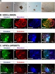
- Figure 7 Characterization of the differentiation ability of hPSCs (hESCs and hiPSCs) in vitro after culture on PVA-oligoVN hydrogels for 20 passages. ( A ) Morphology of EBs differentiated from hESCs (WA09, a,b ) and hiPSCs (HPS0077, c,d ) after culture on PVA-24h-1000 dishes under xeno-free conditions for 20 passages. ( B ) Immunostaining of an ectoderm protein ( a , GFAP; e , betaIII-tubulin), mesoderm protein ( b , SMA), and endoderm ( f , AFP) protein on hESCs (WA09) after culture on PVA-24h-1000 dishes under xeno-free conditions for 20 passages. ( c ) Hoechest staining of hESCs used in ( a,b ). ( d ) Merged picture of ( a-c ). ( g ) Hoechest staining of hESCs used in ( e , f ). (h) Merged picture of ( e-g ). The bar indicates 100 mum. ( C ) Immunostaining of an ectoderm protein ( a , GFAP; e , betaIII-tubulin), mesoderm protein ( b , SMA), and endoderm ( f , AFP) protein on hiPSCs (HPS0077) after culture on PVA-24h-1000 dishes under xeno-free conditions for 20 passages. ( c ) Hoechest staining of hESCs used in ( a,b ). ( d ) Merged picture of ( a-c ). ( g ) Hoechest staining of hESCs used in ( e , f ). ( h ) Merged picture of ( e-g ). The bar indicates 100 mum.
- Submitted by
- Invitrogen Antibodies (provider)
- Main image
- Experimental details
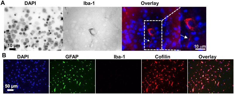
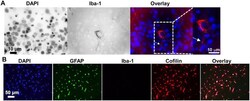
- Fig 3 Microglia cells are virtually absent in dissociated cultures of hippocampal neurons. Microglia and astrocytes in dissociated cultures of mouse hippocampal neurons were characterized by immunoreactivity against Iba-1 or GFAP, respectively. (A) Immunostaining of adult mouse brain slices with Iba-1 antibody revealed the presence of microglia indicating that our fixation, methanol-permeabilization and immunostaining protocol worked well for the Iba-1 antibody. Immunoreactivity is shown in the cytoplasm of a microglial cell co-stained with DAPI (nucleus). A ramified extension of the microglial cell is visible (arrowhead in overlaid image, 100x confocal microscopy). Identical staining was obtained in slices following the more extensive but unnecessary citrate buffer antigen retrieval []. (B) Dissociated cultures of mouse hippocampal neurons (DIV 7) were fixed, methanol permeabilized and immunostained (identical conditions as in panel A). Confocal images were acquired (20x) to reveal nuclei (DAPI), astrocytes (GFAP), microglia (Iba-1), and cofilin and an overlay image generated. No Iba-1 staining was observed indicating cultures were devoid of microglia, a finding confirmed by scanning with 60x and 100x objectives as well. GFAP-positive cells (astrocytes) comprised about 40% of total DAPI nuclei within this particular preparation.