Explore
 Validate
 Learn
Learn Western blot
Antibody data
- Antibody Data
- Antigen structure
- References [7]
- Comments [0]
- Validations
- Western blot [5]
- Immunocytochemistry [4]
- Immunohistochemistry [2]
- Other assay [5]
Submit
Validation data
Reference
Comment
Report error
- Product number
- PA3-16727 - Provider product page

- Provider
- Invitrogen Antibodies
- Product name
- GFAP Polyclonal Antibody
- Antibody type
- Polyclonal
- Antigen
- Purifed from natural sources
- Description
- This antibody is likely to react with most mammals.
- Reactivity
- Human, Mouse, Rat, Bovine, Chicken/Avian, Guinea Pig, Porcine, Rabbit
- Host
- Rabbit
- Isotype
- IgG
- Vial size
- 50 μL
- Concentration
- Conc. Not Determined
- Storage
- Store at 4°C short term. For long term storage, store at -20°C, avoiding freeze/thaw cycles.
Submitted references The mitochondria-targeted antioxidant MitoQ inhibits memory loss, neuropathology, and extends lifespan in aged 3xTg-AD mice.
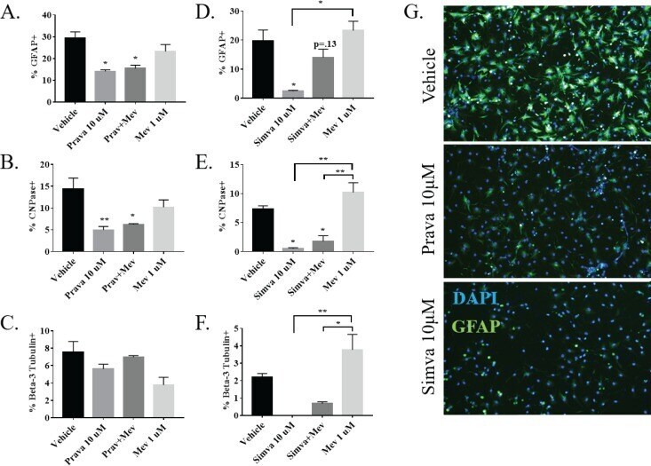
Statins impact primary embryonic mouse neural stem cell survival, cell death, and fate through distinct mechanisms.
Optimizing neuronal differentiation of human pluripotent NT2 stem cells in monolayer cultures.
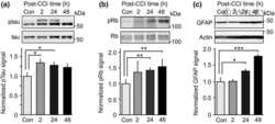
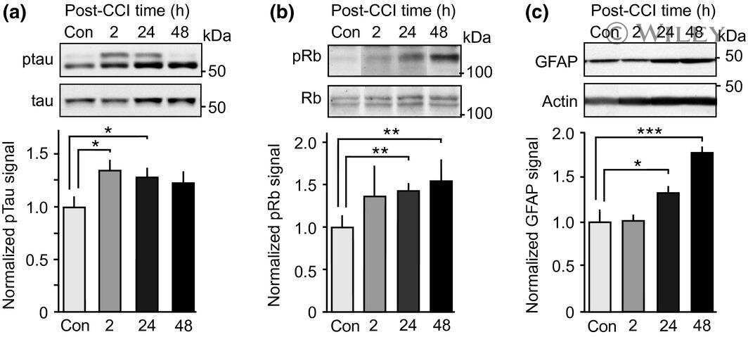
Involvement of aberrant cyclin-dependent kinase 5/p25 activity in experimental traumatic brain injury.
Thioredoxin 1 and glutaredoxin 2 contribute to maintain the phenotype and integrity of neurons following perinatal asphyxia.
Oral administration of the p38α MAPK inhibitor, UR13870, inhibits affective pain behavior after spinal cord injury.
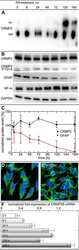
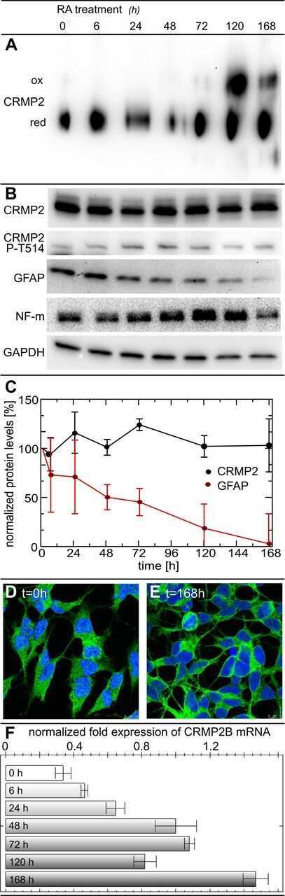
Identification of a dithiol-disulfide switch in collapsin response mediator protein 2 (CRMP2) that is toggled in a model of neuronal differentiation.
Young ML, Franklin JL
Molecular and cellular neurosciences 2019 Dec;101:103409
Molecular and cellular neurosciences 2019 Dec;101:103409
Statins impact primary embryonic mouse neural stem cell survival, cell death, and fate through distinct mechanisms.
Carson RA, Rudine AC, Tally SJ, Franks AL, Frahm KA, Waldman JK, Silswal N, Burale S, Phan JV, Chandran UR, Monaghan AP, DeFranco DB
PloS one 2018;13(5):e0196387
PloS one 2018;13(5):e0196387
Optimizing neuronal differentiation of human pluripotent NT2 stem cells in monolayer cultures.
Abolpour Mofrad S, Kuenzel K, Friedrich O, Gilbert DF
Development, growth & differentiation 2016 Oct;58(8):664-676
Development, growth & differentiation 2016 Oct;58(8):664-676
Involvement of aberrant cyclin-dependent kinase 5/p25 activity in experimental traumatic brain injury.
Yousuf MA, Tan C, Torres-Altoro MI, Lu FM, Plautz E, Zhang S, Takahashi M, Hernandez A, Kernie SG, Plattner F, Bibb JA
Journal of neurochemistry 2016 Jul;138(2):317-27
Journal of neurochemistry 2016 Jul;138(2):317-27
Thioredoxin 1 and glutaredoxin 2 contribute to maintain the phenotype and integrity of neurons following perinatal asphyxia.
Romero JI, Hanschmann EM, Gellert M, Eitner S, Holubiec MI, Blanco-Calvo E, Lillig CH, Capani F
Biochimica et biophysica acta 2015 Jun;1850(6):1274-85
Biochimica et biophysica acta 2015 Jun;1850(6):1274-85
Oral administration of the p38α MAPK inhibitor, UR13870, inhibits affective pain behavior after spinal cord injury.
Galan-Arriero I, Avila-Martin G, Ferrer-Donato A, Gomez-Soriano J, Bravo-Esteban E, Taylor J
Pain 2014 Oct;155(10):2188-98
Pain 2014 Oct;155(10):2188-98
Identification of a dithiol-disulfide switch in collapsin response mediator protein 2 (CRMP2) that is toggled in a model of neuronal differentiation.
Gellert M, Venz S, Mitlöhner J, Cott C, Hanschmann EM, Lillig CH
The Journal of biological chemistry 2013 Dec 6;288(49):35117-25
The Journal of biological chemistry 2013 Dec 6;288(49):35117-25
No comments: Submit comment
Supportive validation
- Submitted by
- Invitrogen Antibodies (provider)
- Main image
- Experimental details
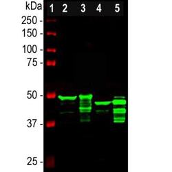
- Western blot analysis of GFAP in different tissue lysates. Samples were incubated in GFAP polyclonal antibody (Product # PA3-16727 using a dilution of 1:5000. Antibody in green: [1] protein standard (red), [2] rat brain, [3] rat spinal cord, [4] mouse brain, [5] mouse spinal cord. Strong band at about 50 kDa corresponds to the major isotype of the GFAP protein. Smaller isotypes and proteolytic fragments of GFAP are also detected on the blot.

- Submitted by
- Invitrogen Antibodies (provider)
- Main image
- Experimental details
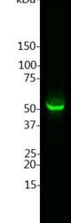
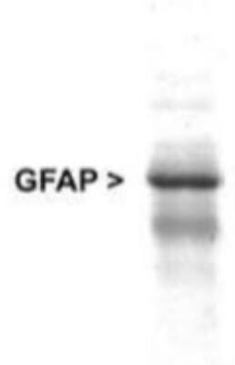
- Western blot analysis of GFAP in rat brain lysate. Samples were incubated in GFAP polyclonal antibody (Product # PA3-16727 using a dilution of 1:5000. Specific band running with an apparent SDS-PAGE molecular weight of ~50 kDa corresponds to rodent GFAP was observed.
- Submitted by
- Invitrogen Antibodies (provider)
- Main image

- Experimental details
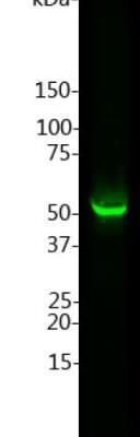
- Western blot analysis of GFAP in whole rat cerebellum homogenate. Sample was incubated in GFAP polyclonal antibody (Product # PA3-16727).
- Submitted by
- Invitrogen Antibodies (provider)
- Main image

- Experimental details
- Western blot analysis of GFAP in 0.05 mg/mL Human Brain lysate. Samples were incubated in GFAP polyclonal antibody (Product # PA3-16727). This experiment was performed under reducing conditions using the 12-230 kDa separation system.

- Submitted by
- Invitrogen Antibodies (provider)
- Main image

- Experimental details
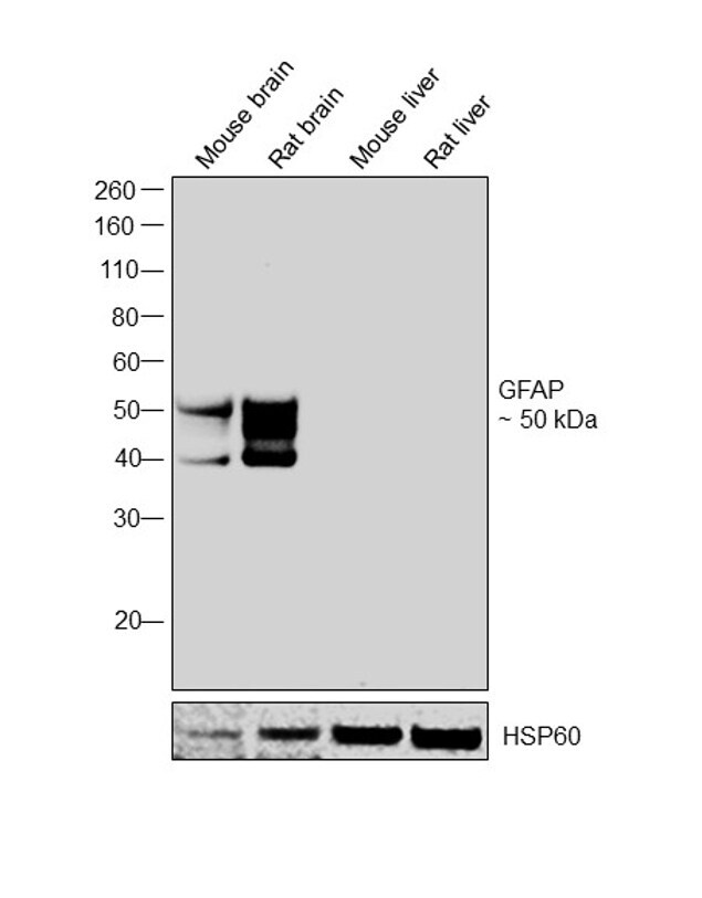
- Western blot was performed using Anti-GFAP Rabbit Polyclonal Antibody (Product # PA3-16727) and a 50 kDa band corresponding to GFAP was observed across tissues tested except Mouse and Rat liver. Whole cell extracts (30 µg lysate) of Mouse brain (Lane 1), Rat brain (Lane 2), Mouse liver (Lane 3) and Rat liver (Lane 4) were electrophoresed using Novex® NuPAGE® 4-12 % Bis-Tris gel (Product # NP0321BOX). Resolved proteins were then transferred onto a nitrocellulose membrane (Product # IB23001) by iBlot® 2 Dry Blotting System (Product # IB21001). The blot was probed with the primary antibody (1:20000 dilution) and detected by chemiluminescence with Goat anti-Rabbit IgG (Heavy Chain), Superclonal™ Recombinant Secondary Antibody, HRP (Product # A27036, 1:4000 dilution) using the iBright FL 1000 (Product # A32752). Chemiluminescent detection was performed using Novex® ECL Chemiluminescent Substrate Reagent Kit (Product # WP20005).
Supportive validation
- Submitted by
- Invitrogen Antibodies (provider)
- Main image

- Experimental details
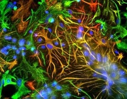
- Immunocytochemistry analysis of GFAP in mixed neuron-glial cultures. Samples were incubated in GFAP polyclonal antibody (Product # PA3-16727). GFAP antibody (red) and Vimentin antibody (green). The fibroblastic cells contain only Vimentin and so are green. The astrocytes contain either Vimentin and GFAP (appearing golden) or predominantly GFAP (appearing red). Blue is nuclear DNA stain.

- Submitted by
- Invitrogen Antibodies (provider)
- Main image

- Experimental details
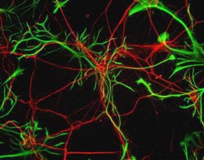
- Immunocytochemistry analysis of GFAP in Rat neurons. Samples were incubated in GFAP polyclonal antibody (Product # PA3-16727). Neurofilament Heavy antibody (red) and GFAP antibody (green).

- Submitted by
- Invitrogen Antibodies (provider)
- Main image
- Experimental details
- Immunocytochemistry analysis of GFAP in mixed neuron-glial cultures. Samples were incubated in GFAP polyclonal antibody (Product # PA3-16727). GFAP antibody (red) and Vimentin antibody (green). The fibroblastic cells contain only Vimentin and so are green. The astrocytes contain either Vimentin and GFAP (appearing golden) or predominantly GFAP (appearing red). Blue is nuclear DNA stain.

- Submitted by
- Invitrogen Antibodies (provider)
- Main image

- Experimental details
- Immunocytochemistry analysis of GFAP in Rat neurons. Samples were incubated in GFAP polyclonal antibody (Product # PA3-16727). Neurofilament Heavy antibody (red) and GFAP antibody (green).
Supportive validation
- Submitted by
- Invitrogen Antibodies (provider)
- Main image
- Experimental details
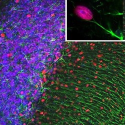
- Immunohistochemical analysis of GFAP in rat cerebellum section. Samples were incubated in GFAP polyclonal antibody (Product # PA3-16727) using a dilution of 1:5000. Antibody in green and mouse monoclonal antibody to MeCP2, dilution 1:500, in red. The blue is DAPI staining of nuclear DNA. Following transcardial perfusion of rat with 4% paraformaldehyde, brain was post fixed for 1 hour, cut to 45 µM, and free-floating sections were stained with above antibodies. The GFAP antibody stains the network of astrocytic cells and the processes of Bergmann glia in the molecular layer. The MeCP2 antibody specifically labels nuclei of certain neurons.

- Submitted by
- Invitrogen Antibodies (provider)
- Main image
- Experimental details
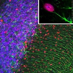
- Immunohistochemical analysis of GFAP in rat cerebellum section. Samples were incubated in GFAP polyclonal antibody (Product # PA3-16727) using a dilution of 1:5000. Antibody in green and mouse monoclonal antibody to MeCP2, dilution 1:500, in red. The blue is DAPI staining of nuclear DNA. Following transcardial perfusion of rat with 4% paraformaldehyde, brain was post fixed for 1 hour, cut to 45 µM, and free-floating sections were stained with above antibodies. The GFAP antibody stains the network of astrocytic cells and the processes of Bergmann glia in the molecular layer. The MeCP2 antibody specifically labels nuclei of certain neurons.

Supportive validation
- Submitted by
- Invitrogen Antibodies (provider)
- Main image
- Experimental details
- NULL
- Submitted by
- Invitrogen Antibodies (provider)
- Main image
- Experimental details
- NULL
- Submitted by
- Invitrogen Antibodies (provider)
- Main image
- Experimental details
- NULL
- Submitted by
- Invitrogen Antibodies (provider)
- Main image
- Experimental details
- NULL
- Submitted by
- Invitrogen Antibodies (provider)
- Main image
- Experimental details
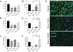
- Fig 7 Statins differentially alter NSPC fate through a non-CBP dependent mechanism. Percentage of (A) GFAP+ glia (n = 3), (B) CNPase+ oligodendrocytes (n = 3), and (C) Beta-3 Tubulin+ neurons (n = 3) present in a population of NSPCs differentiated for three days in the presence of vehicle, 10 muM prava, 1 muM mevalonate (mev), or prava and mev. (D-F) The same experiment performed with 5muM simvastatin. (G) Representative images of differentiated cells stained with DAPI and GFAP. (*p