Antibody data
- Antibody Data
- Antigen structure
- References [13]
- Comments [0]
- Validations
- Western blot [2]
- Immunocytochemistry [1]
- Immunohistochemistry [1]
Submit
Validation data
Reference
Comment
Report error
- Product number
- MA5-14084 - Provider product page
- Provider
- Invitrogen Antibodies
- Product name
- Actin Muscle Monoclonal Antibody (HHF35)
- Antibody type
- Monoclonal
- Antigen
- Other
- Description
- This antibody reacts with actin from tissue extracts of uterus, ileum, aorta, diaphragm, heart and smooth muscle cells. It recognizes alpha-actin from skeletal, cardiac, and smooth muscle, and reacts with gamma actin from smooth muscle. This antibody will react with tumors from smooth muscle (leiomyomas and leiomyosarcomas) as well as skeletal muscle tumors (rhabdomyomas and rhabdomyosarcomas).
- Antibody clone number
- HHF35
- Concentration
- Conc. Not Determined
Submitted references Evidence for CD34/SMA positive cells in the left main coronary artery in atherogenesis.
Plexiform fibromyxoma (plexiform angiomyxoid myofibroblastic tumor) of stomach: an unusual presentation as a fistulating abscess.
Expression of E-cadherin in angiomyolipoma.
Sporadic haemangioblastoma of the kidney with rhabdoid features and focal CD10 expression: report of a case and literature review.
Fascin expression and its potential significance in gastrointestinal stromal tumors.
Tumor-in-tumor of the thyroid with basaloid differentiation: a lesion with a solid cell nest neoplastic component?
G-CSF promotes the proliferation of developing cardiomyocytes in vivo and in derivation from ESCs and iPSCs.
Chorangiosis: the potential role of smoking and air pollution.
Myointimoma of the glans penis.
Microfabrication-based modulation of embryonic stem cell differentiation.
Cutaneous lymphoid hyperplasia and other lymphoid infiltrates of the breast nipple: a retrospective clinicopathologic study of fifty-six patients.
Ectopic cervical anaplastic ependymoma.
Epstein-Barr virus-associated smooth muscle tumour: a distinctive mesenchymal tumour of immunocompromised individuals.
Kruzliak P, Hare DL, Sabaka P, Delev D, Gaspar L, Rodrigo L, Zulli A
Acta histochemica 2016 May;118(4):413-7
Plexiform fibromyxoma (plexiform angiomyxoid myofibroblastic tumor) of stomach: an unusual presentation as a fistulating abscess.
Lee PW, Yau DT, Lau PP, Chan JK
International journal of surgical pathology 2014 May;22(3):286-90
Expression of E-cadherin in angiomyolipoma.
Wang Z, Gong Q, Fan Q
Human pathology 2012 Dec;43(12):2348-53
Sporadic haemangioblastoma of the kidney with rhabdoid features and focal CD10 expression: report of a case and literature review.
Yin WH, Li J, Chan JK
Diagnostic pathology 2012 Apr 12;7:39
Fascin expression and its potential significance in gastrointestinal stromal tumors.
Ozcan A, Karslioğlu Y, Günal A, Cermık AH, Kurt B, Ongürü O
The Turkish journal of gastroenterology : the official journal of Turkish Society of Gastroenterology 2011 Aug;22(4):363-8
Tumor-in-tumor of the thyroid with basaloid differentiation: a lesion with a solid cell nest neoplastic component?
Eloy C, Vinagre J, Cameselle-Teijeiro J, Paiva ME, Soares P, Sobrinho-Simões M
International journal of surgical pathology 2011 Apr;19(2):276-80
G-CSF promotes the proliferation of developing cardiomyocytes in vivo and in derivation from ESCs and iPSCs.
Shimoji K, Yuasa S, Onizuka T, Hattori F, Tanaka T, Hara M, Ohno Y, Chen H, Egasgira T, Seki T, Yae K, Koshimizu U, Ogawa S, Fukuda K
Cell stem cell 2010 Mar 5;6(3):227-37
Chorangiosis: the potential role of smoking and air pollution.
Akbulut M, Sorkun HC, Bir F, Eralp A, Duzcan E
Pathology, research and practice 2009;205(2):75-81
Myointimoma of the glans penis.
Vardar E, Gunlusoy B, Arslan M, Kececi S
Pathology international 2007 Mar;57(3):158-61
Microfabrication-based modulation of embryonic stem cell differentiation.
Park J, Cho CH, Parashurama N, Li Y, Berthiaume F, Toner M, Tilles AW, Yarmush ML
Lab on a chip 2007 Aug;7(8):1018-28
Cutaneous lymphoid hyperplasia and other lymphoid infiltrates of the breast nipple: a retrospective clinicopathologic study of fifty-six patients.
Boudova L, Kazakov DV, Sima R, Vanecek T, Torlakovic E, Lamovec J, Kutzner H, Szepe P, Plank L, Bouda J, Hes O, Mukensnabl P, Michal M
The American Journal of dermatopathology 2005 Oct;27(5):375-86
Ectopic cervical anaplastic ependymoma.
Wang Z, Huang G, Yan P, Liang R, Wang J, Yan Q, Zhang J, Cheng H, Hu P, Ma MJ
Pathology international 2005 Dec;55(12):781-4
Epstein-Barr virus-associated smooth muscle tumour: a distinctive mesenchymal tumour of immunocompromised individuals.
Cheuk W, Li PC, Chan JK
Pathology 2002 Jun;34(3):245-9
No comments: Submit comment
Supportive validation
- Submitted by
- Invitrogen Antibodies (provider)
- Main image
- Experimental details
- Western blot analysis of Actin Muscle Specific was performed by loading 25 µg of mouse skeletal muscle (Lane 1), Raji (Lane 2) and Hela (Lane 3) cell lysates onto an SDS polyacrylamide gel. Proteins were transferred to a PVDF membrane and blocked at 4ºC overnight. The membrane was probed with a Actin Muscle Specific monoclonal antibody (Product # MA5-14084) at a dilution of 1:500 (mouse) and 1:100 (Raji and Hela) overnight at 4°C, washed in TBST, and probed with an HRP-conjugated secondary antibody for 1 hr at room temperature in the dark. Chemiluminescent detection was performed using Pierce ECL Plus Western Blotting Substrate (Product # 32132). Results show a band at approx. 42 kDa.
- Submitted by
- Invitrogen Antibodies (provider)
- Main image
- Experimental details
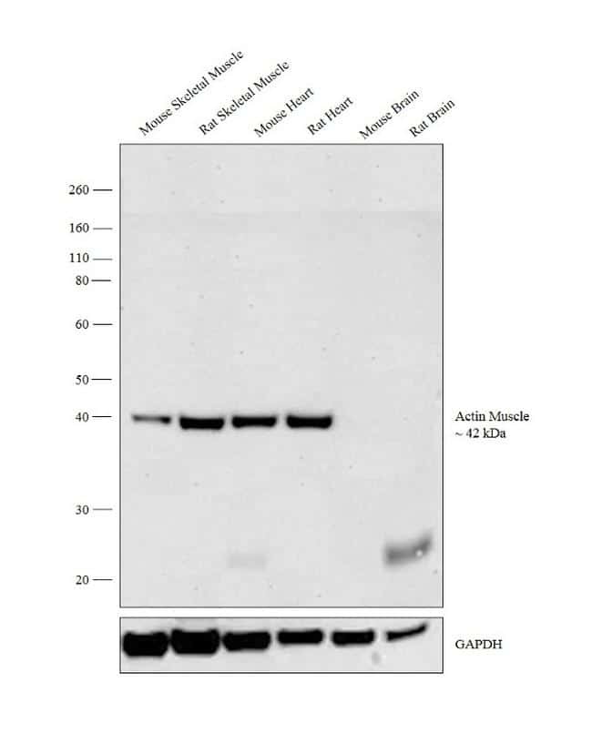
- Western blot analysis was performed on tissue extracts (30 µg lysate) of Mouse Skeletal Muscle (Lane 1), Rat Skeletal Muscle (Lane 2), Mouse Heart (Lane 3), Rat Heart (Lane 4), Mouse Brain (Lane 5) and Rat Brain (Lane 6). The blot was probed with Anti-Actin Muscle Monoclonal Antibody (Product # MA5-14084, 1:500 dilution) and detected by chemiluminescence using Goat anti-Mouse IgG (H+L) Superclonal™ Secondary Antibody, HRP conjugate (Product # A28177, 0.25 µg/ml, 1:4000 dilution). A 42 kDa band corresponding to Actin Muscle was observed across the muscle tissues tested and not in non muscle tissues.
Supportive validation
- Submitted by
- Invitrogen Antibodies (provider)
- Main image
- Experimental details
- Immunofluorescent analysis of Actin Muscle Specific (green) showing staining in the cytoplasm of Hela cells (right) compared to a negative control without primary antibody (left). Formalin-fixed cells were permeabilized with 0.1% Triton X-100 in TBS for 5-10 minutes and blocked with 3% BSA-PBS for 30 minutes at room temperature. Cells were probed with an Actin Muscle Specific monoclonal antibody (Product # MA5-14084) in 3% BSA-PBS at a dilution of 1:20 and incubated overnight at 4ºC in a humidified chamber. Cells were washed with PBST and incubated with a DyLight-conjugated secondary antibody in PBS at room temperature in the dark. F-actin (red) was stained with a fluorescent red phalloidin and nuclei (blue) were stained with Hoechst or DAPI. Images were taken at a magnification of 100x.
Supportive validation
- Submitted by
- Invitrogen Antibodies (provider)
- Main image
- Experimental details
- Formalin-fixed, paraffin-embedded human skeletal Muscle stained with Muscle Specific Actin antibody using peroxidase-conjugate and DAB chromogen. Note cytoplasmic staining of muscle cells.