Explore
 Validate
 Learn
LearnNB600-985
antibody from Novus Biologicals
Targeting: CD68
DKFZp686M18236, GP110, LAMP4, macrosialin, SCARD1
 Western blot Immunocytochemistry Immunoprecipitation
 Immunohistochemistry Flow cytometry Radioimmunoassay
Antibody data
- Antibody Data
- Antigen structure
- References [6]
- Comments [0]
- Validations
- Immunohistochemistry [3]
- Flow cytometry [7]
Submit
Validation data
Reference
Comment
Report error
- Product number
- NB600-985 - Provider product page

- Provider
- Novus Biologicals
- Proper citation
- Novus Cat#NB600-985, RRID:AB_791231
- Product name
- Mouse Monoclonal CD68/SR-D1 Antibody
- Antibody type
- Monoclonal
- Description
- Protein A or G purified. NB600-985 recognizes a single chain glycoprotein of 110kD that is expressed predominantly on the lysosomal membrane of myeloid cells. Weak cell surface expression also occurs. The antigen is expressed by the majority of tissue macrophages and weakly by peripheral blood granulocytes. Studies have shown that the antigen recognized by ED1 is the rat homologue of human CD68.
- Reactivity
- Human, Mouse, Rat, Bovine, Feline, Simian
- Host
- Mouse
- Isotype
- IgG
- Vial size
- 0.125 mg
- Concentration
- 1.0 mg/ml
- Storage
- Store at 4C short term. Aliquot and store at -20C long term. Avoid freeze-thaw cycles.
Submitted references Revisiting the "race for the surface" in a pre-clinical model of implant infection.
Site-Specific Reprogramming of Macrophage Responsiveness to Bacterial Lipopolysaccharide in Obesity.
Crystal deposition triggers tubule dilation that accelerates cystogenesis in polycystic kidney disease.
CEST MRI of sepsis-induced acute kidney injury.
CorMatrix Wrapped Around the Adventitia of the Arteriovenous Fistula Outflow Vein Attenuates Venous Neointimal Hyperplasia.
Autologous minced muscle grafts improve endogenous fracture healing and muscle strength after musculoskeletal trauma.
Shiels SM, Mangum LH, Wenke JC
European cells & materials 2020 Jan 29;39:77-95
European cells & materials 2020 Jan 29;39:77-95
Site-Specific Reprogramming of Macrophage Responsiveness to Bacterial Lipopolysaccharide in Obesity.
Komegae EN, Fonseca MT, da Silveira Cruz-Machado S, Turato WM, Filgueiras LR, Markus RP, Steiner AA
Frontiers in immunology 2019;10:1496
Frontiers in immunology 2019;10:1496
Crystal deposition triggers tubule dilation that accelerates cystogenesis in polycystic kidney disease.
Torres JA, Rezaei M, Broderick C, Lin L, Wang X, Hoppe B, Cowley BD Jr, Savica V, Torres VE, Khan S, Holmes RP, Mrug M, Weimbs T
The Journal of clinical investigation 2019 Jul 30;129(10):4506-4522
The Journal of clinical investigation 2019 Jul 30;129(10):4506-4522
CEST MRI of sepsis-induced acute kidney injury.
Liu J, Han Z, Chen G, Li Y, Zhang J, Xu J, van Zijl PCM, Zhang S, Liu G
NMR in biomedicine 2018 Aug;31(8):e3942
NMR in biomedicine 2018 Aug;31(8):e3942
CorMatrix Wrapped Around the Adventitia of the Arteriovenous Fistula Outflow Vein Attenuates Venous Neointimal Hyperplasia.
Yang B, Kilari S, Brahmbhatt A, McCall DL, Torres EN, Leof EB, Mukhopadhyay D, Misra S
Scientific reports 2017 Oct 30;7(1):14298
Scientific reports 2017 Oct 30;7(1):14298
Autologous minced muscle grafts improve endogenous fracture healing and muscle strength after musculoskeletal trauma.
Hurtgen BJ, Ward CL, Leopold Wager CM, Garg K, Goldman SM, Henderson BEP, McKinley TO, Greising SM, Wenke JC, Corona BT
Physiological reports 2017 Jul;5(14)
Physiological reports 2017 Jul;5(14)
No comments: Submit comment
Supportive validation
- Submitted by
- Novus Biologicals (provider)
- Main image

- Experimental details
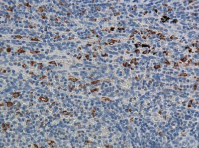
- Immunohistochemistry-Paraffin: CD68/SR-D1 Antibody (ED1) [NB600-985] - Cynomolgus monkey spleen. Image from verified customer review.
- Submitted by
- Novus Biologicals (provider)
- Main image
- Experimental details
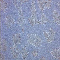
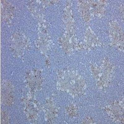
- Immunohistochemistry-Paraffin: CD68/SR-D1 Antibody (ED1) [NB600-985] - Staining of rat liver
- Submitted by
- Novus Biologicals (provider)
- Main image
- Experimental details
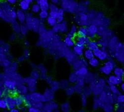
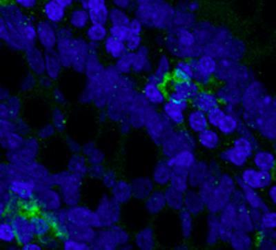
- Immunohistochemistry-Frozen: CD68/SR-D1 Antibody (ED1) [NB600-985] - The feline macrophage containing section was incubated with the antibody at 1:50 and the section was further stained with Alexa secondary antibody. Image was captured with an epifluorescence microscope.This image was submitted via customer review.
Supportive validation
- Submitted by
- Novus Biologicals (provider)
- Main image

- Experimental details
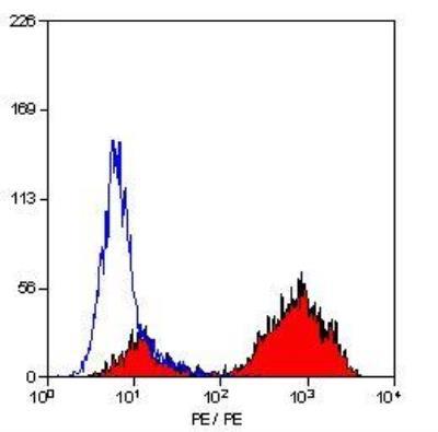
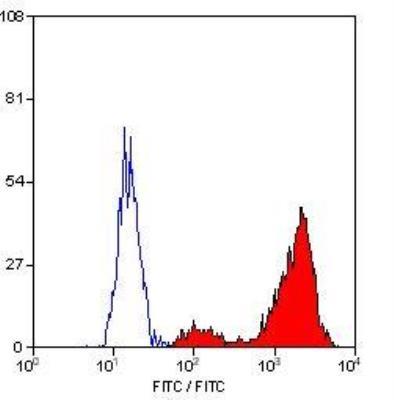
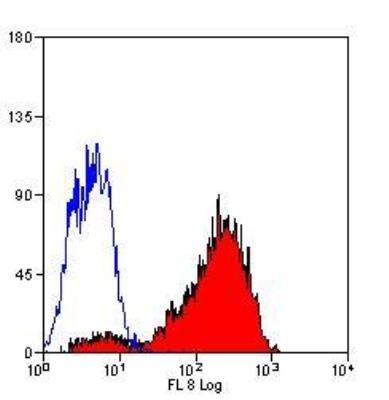
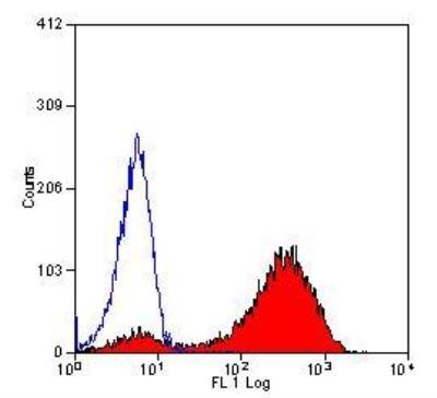
- Flow Cytometry: CD68/SR-D1 Antibody (ED1) [NB600-985] - Staining of rat peritoneal macrophages cells.
- Submitted by
- Novus Biologicals (provider)
- Main image

- Experimental details
- Flow Cytometry: CD68/SR-D1 Antibody (ED1) [NB600-985] - Staining of rat peritoneal macrophages cells.
- Submitted by
- Novus Biologicals (provider)
- Main image

- Experimental details
- Flow Cytometry: CD68/SR-D1 Antibody (ED1) [NB600-985] - Staining of rat peritoneal macrophages.
- Submitted by
- Novus Biologicals (provider)
- Main image

- Experimental details
- Flow Cytometry: CD68/SR-D1 Antibody (ED1) [NB600-985] - Staining of rat peritoneal macrophages
- Submitted by
- Novus Biologicals (provider)
- Main image
- Experimental details
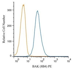
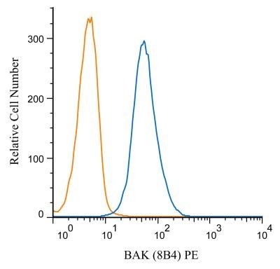
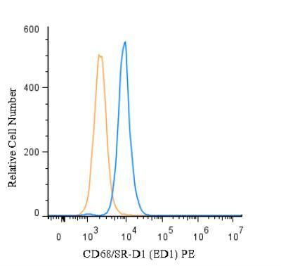
- Flow Cytometry: CD68/SR-D1 Antibody (ED1) [NB600-985] - An intracellular stain was performed on Jurkat cells with BAK antibody (8B4) NBP1-74026PE (blue) and a matched isotype control NB600-985PE (orange). Cells were fixed with 4% PFA and then permeablized with 0.1% saponin. Cells were incubated in an antibody dilution of 1 ug/mL for 30 minutes at room temperature. Both antibodies were conjugated to phycoerythrin. Image using the PE form of this antibody.
- Submitted by
- Novus Biologicals (provider)
- Main image

- Experimental details
- Flow (Intracellular): CD68/SR-D1 Antibody (ED1) [NB600-985] - An intracellular stain was performed on U-937 cells with CD68/SR-D1 Antibody (ED1) NB600-985PE (blue) and a matched isotype control (orange). Cells were fixed with 4% PFA and then permeabilized with 0.1% saponin. Cells were incubated in an antibody dilution of 5 ug/mL for 30 minutes at room temperature. Both antibodies were conjugated to phycoerythrin.
- Submitted by
- Novus Biologicals (provider)
- Main image

- Experimental details
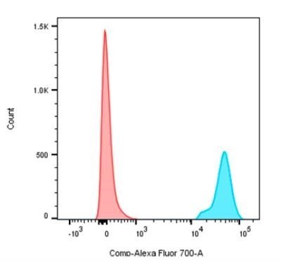
- Flow Cytometry: CD68/SR-D1 Antibody (ED1) [NB600-985] - Decidual Macrophages were analyzed with a BD LSRFortessa. Antibody was diluted 1:100 in staining buffer before use. Isotype control is in Red. Sample with NB600-985AF700 is in Blue. Image from the Alexa Fluor 700 version of this antibody.