 Explore
 Validate
 Learn
Learn Immunocytochemistry
Antibody data
- Antibody Data
- Antigen structure
- References [1]
- Comments [0]
- Validations
- Immunocytochemistry [1]
Submit
Validation data
Reference
Comment
Report error
- Product number
- MA1-82873 - Provider product page

- Provider
- Invitrogen Antibodies
- Product name
- Lysozyme Monoclonal Antibody (BGN/0696/5B1)
- Antibody type
- Monoclonal
- Antigen
- Other
- Description
- Heat-mediated antigen retrieval is recommended for the staining of paraffin sections. A suggested positive control for this product is human tonsil. For FACS analysis, use 10 µL of the suggested working dilution to label 1x10^6 cells in 100 µL.
- Antibody clone number
- BGN/0696/5B1
- Concentration
- 1 mg/mL
Submitted references Direct On-Chip Differentiation of Intestinal Tubules from Induced Pluripotent Stem Cells.
Naumovska E, Aalderink G, Wong Valencia C, Kosim K, Nicolas A, Brown S, Vulto P, Erdmann KS, Kurek D
International journal of molecular sciences 2020 Jul 14;21(14)
International journal of molecular sciences 2020 Jul 14;21(14)
No comments: Submit comment
Supportive validation
- Submitted by
- Invitrogen Antibodies (provider)
- Main image
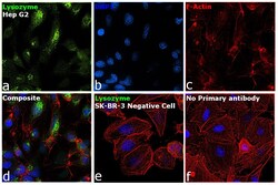
- Experimental details
- Immunofluorescence analysis of Lysozyme was performed using 70% confluent log phase Hep G2 cells. The cells were fixed with 4% paraformaldehyde for 10 minutes, permeabilized with 0.1% Triton™ X-100 for 15 minutes, and blocked with 2% BSA for 1 hour at room temperature. The cells were labeled with Lysozyme Mouse Monoclonal Antibody (BGN/0696/5B1) (Product # MA1-82873) at 1:100 dilution in 0.1% BSA, incubated at 4 degree Celsius overnight and then labeled with Goat anti-Mouse IgG (H+L), Superclonal™ Recombinant Secondary Antibody, Alexa Fluor 488 conjugate (Product # A28175) at a dilution of 1:2000 for 45 minutes at room temperature (Panel a: green). Nuclei (Panel b: blue) were stained with SlowFade® Gold Antifade Mountant with DAPI (Product # S36938). F-actin (Panel c: red) was stained with Rhodamine Phalloidin (Product # R415, 1:300). Panel d represents the merged image showing localization to golgi network and cytoplasm. Panel e shows SK-BR-3 cells with no expression of Lysozyme. Panel f represents control cells with no primary antibody to assess background. The images were captured at 60X magnification.
