Explore
 Validate
 Learn
Learn Western blot
 Immunocytochemistry
Antibody data
- Antibody Data
- Antigen structure
- References [7]
- Comments [0]
- Validations
- Immunocytochemistry [2]
- Immunohistochemistry [1]
- Other assay [2]
Submit
Validation data
Reference
Comment
Report error
- Product number
- PA1-29680 - Provider product page

- Provider
- Invitrogen Antibodies
- Product name
- Lysozyme Polyclonal Antibody
- Antibody type
- Polyclonal
- Antigen
- Other
- Description
- Predigestion with a proteolytic enzyme improves staining of formalin fixed paraffin embedded tissue sections.
- Reactivity
- Human
- Host
- Rabbit
- Isotype
- IgG
- Vial size
- 500 μL
- Storage
- 4°C
Submitted references A potent HNF4α agonist reveals that HNF4α controls genes important in inflammatory bowel disease and Paneth cells.
The Effect of Thiol Structure on Allyl Sulfide Photodegradable Hydrogels and their Application as a Degradable Scaffold for Organoid Passaging.
Relaxation of Extracellular Matrix Forces Directs Crypt Formation and Architecture in Intestinal Organoids.
Impact of Maternal Malnutrition on Gut Barrier Defense: Implications for Pregnancy Health and Fetal Development.
Ret receptor tyrosine kinase sustains proliferation and tissue maturation in intestinal epithelia.
Kaiso differentially regulates components of the Notch signaling pathway in intestinal cells.
Paneth cell-mediated multiorgan dysfunction after acute kidney injury.
Lee SH, Veeriah V, Levine F
PloS one 2022;17(4):e0266066
PloS one 2022;17(4):e0266066
The Effect of Thiol Structure on Allyl Sulfide Photodegradable Hydrogels and their Application as a Degradable Scaffold for Organoid Passaging.
Yavitt FM, Brown TE, Hushka EA, Brown ME, Gjorevski N, Dempsey PJ, Lutolf MP, Anseth KS
Advanced materials (Deerfield Beach, Fla.) 2020 Jul;32(30):e1905366
Advanced materials (Deerfield Beach, Fla.) 2020 Jul;32(30):e1905366
Relaxation of Extracellular Matrix Forces Directs Crypt Formation and Architecture in Intestinal Organoids.
Hushka EA, Yavitt FM, Brown TE, Dempsey PJ, Anseth KS
Advanced healthcare materials 2020 Apr;9(8):e1901214
Advanced healthcare materials 2020 Apr;9(8):e1901214
Impact of Maternal Malnutrition on Gut Barrier Defense: Implications for Pregnancy Health and Fetal Development.
Srugo SA, Bloise E, Nguyen TTN, Connor KL
Nutrients 2019 Jun 19;11(6)
Nutrients 2019 Jun 19;11(6)
Ret receptor tyrosine kinase sustains proliferation and tissue maturation in intestinal epithelia.
Perea D, Guiu J, Hudry B, Konstantinidou C, Milona A, Hadjieconomou D, Carroll T, Hoyer N, Natarajan D, Kallijärvi J, Walker JA, Soba P, Thapar N, Burns AJ, Jensen KB, Miguel-Aliaga I
The EMBO journal 2017 Oct 16;36(20):3029-3045
The EMBO journal 2017 Oct 16;36(20):3029-3045
Kaiso differentially regulates components of the Notch signaling pathway in intestinal cells.
Robinson SC, Klobucar K, Pierre CC, Ansari A, Zhenilo S, Prokhortchouk E, Daniel JM
Cell communication and signaling : CCS 2017 Jun 21;15(1):24
Cell communication and signaling : CCS 2017 Jun 21;15(1):24
Paneth cell-mediated multiorgan dysfunction after acute kidney injury.
Park SW, Kim M, Kim JY, Ham A, Brown KM, Mori-Akiyama Y, Ouellette AJ, D'Agati VD, Lee HT
Journal of immunology (Baltimore, Md. : 1950) 2012 Dec 1;189(11):5421-33
Journal of immunology (Baltimore, Md. : 1950) 2012 Dec 1;189(11):5421-33
No comments: Submit comment
Supportive validation
- Submitted by
- Invitrogen Antibodies (provider)
- Main image
- Experimental details
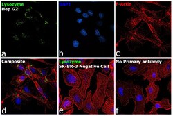
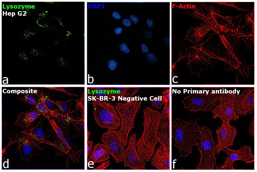
- Immunofluorescence analysis of Lysozyme was performed using 70% confluent log phase Hep G2 cells. The cells were fixed with 4% paraformaldehyde for 10 minutes, permeabilized with 0.1% Triton™ X-100 for 15 minutes, and blocked with 2% BSA for 1 hour at room temperature. The cells were labeled with Lysozyme Rabbit Polyclonal Antibody (Product # PA1-29680) at 1:100 dilution in 0.1% BSA, incubated at 4 degree Celsius overnight and then labeled with Goat anti-Rabbit IgG (H+L) Superclonal™ Recombinant Secondary Antibody, Alexa Fluor® 488 conjugate (Product # A27034) at a dilution of 1:2000 for 45 minutes at room temperature (Panel a: green). Nuclei (Panel b: blue) were stained with SlowFade® Gold Antifade Mountant with DAPI (Product # S36938). F-actin (Panel c: red) was stained with Rhodamine Phalloidin (Product # R415, 1:300). Panel d represents the merged image showing localization to golgi network and cytoplasm. Panel e shows SK-BR-3 cells with no expression of Lysozyme. Panel f represents control cells with no primary antibody to assess background. The images were captured at 60X magnification.
- Submitted by
- Invitrogen Antibodies (provider)
- Main image

- Experimental details
- Immunofluorescence analysis of Lysozyme was performed using 70% confluent log phase Hep G2 cells. The cells were fixed with 4% paraformaldehyde for 10 minutes, permeabilized with 0.1% Triton™ X-100 for 15 minutes, and blocked with 2% BSA for 1 hour at room temperature. The cells were labeled with Lysozyme Rabbit Polyclonal Antibody (Product # PA1-29680) at 1:100 dilution in 0.1% BSA, incubated at 4 degree Celsius overnight and then labeled with Goat anti-Rabbit IgG (Heavy Chain) Superclonal™ Recombinant Secondary Antibody, Alexa Fluor® 488 conjugate (Product # A27034) at a dilution of 1:2000 for 45 minutes at room temperature (Panel a: green). Nuclei (Panel b: blue) were stained with SlowFade® Gold Antifade Mountant with DAPI (Product # S36938). F-actin (Panel c: red) was stained with Rhodamine Phalloidin (Product # R415, 1:300). Panel d represents the merged image showing localization to golgi network and cytoplasm. Panel e shows SK-BR-3 cells with no expression of Lysozyme. Panel f represents control cells with no primary antibody to assess background. The images were captured at 60X magnification.

Supportive validation
- Submitted by
- Invitrogen Antibodies (provider)
- Main image
- Experimental details
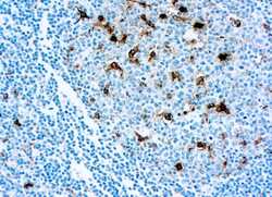
- Immunohistochemical (Paraffin) analysis of human tonsil tissue using (Product # PA1-29680) Lysozyme Polyclonal Antibody.

Supportive validation
- Submitted by
- Invitrogen Antibodies (provider)
- Main image
- Experimental details
- NULL
- Submitted by
- Invitrogen Antibodies (provider)
- Main image
- Experimental details
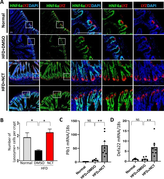
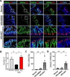
- 10.1371/journal.pone.0266066.g004 Fig 4 NCT induced recovery of Paneth cells in intestine. A . Frozen sections of intestine from the mice described in Fig 1 were stained with HNF4alpha (green), Lysozyme (red) and DAPI (blue) in mice fed normal diet, HFD+DMSO or HFD+NCT. White box indicates high power view in adjacent panel. B. Quantification of the number of lysozyme-positive cells per intestinal crypt (HFD+DMSO vs. Normal or HFD+NCT). C, D . qPCR analysis in mouse small intestine of Plb1 and Defa22 mRNA expression normalized with 18s rRNA (N = 6-8). NS = non-significant, * p