Explore
 Validate
 Learn
Learn Western blot
 Immunocytochemistry
Antibody data
- Antibody Data
- Antigen structure
- References [1]
- Comments [0]
- Validations
- Western blot [2]
- Immunohistochemistry [1]
- Flow cytometry [1]
Submit
Validation data
Reference
Comment
Report error
- Product number
- NBP2-61118 - Provider product page

- Provider
- Novus Biologicals
- Product name
- Rabbit Polyclonal Lysozyme Antibody
- Antibody type
- Polyclonal
- Description
- Immunogen affinity purified.
- Reactivity
- Human, Mouse
- Host
- Rabbit
- Isotype
- IgG
- Vial size
- 0.1 mg
- Concentration
- 1.0 mg/ml
- Storage
- Store at 4C short term. Aliquot and store at -20C long term. Avoid freeze-thaw cycles.
Submitted references The Misshapen subfamily of Ste20 kinases regulate proliferation in the aging mammalian intestinal epithelium.
Li Q, Nirala NK, Chen HJ, Nie Y, Wang W, Zhang B, Czech MP, Wang Q, Xu L, Mao J, Ip YT
Journal of cellular physiology 2019 Dec;234(12):21925-21936
Journal of cellular physiology 2019 Dec;234(12):21925-21936
No comments: Submit comment
Supportive validation
- Submitted by
- Novus Biologicals (provider)
- Main image

- Experimental details
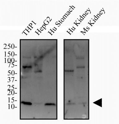
- Western Blot: Lysozyme Antibody [NBP2-61118] - Total protein from human cell lines THP-1 and HepG2, human stomach and kidney as well as mouse kidney was separated on a 4-20% gel by SDS-PAGE, transferred to 0.2 um PVDF membrane and blocked in 5% non-fat milk in TBST. The membrane was probed with 2.0 ug/ml anti-Lysozyme in 5% non-fat milk in TBST and detected with an anti-rabbit HRP secondary antibody using chemiluminescence.
- Submitted by
- Novus Biologicals (provider)
- Main image
- Experimental details
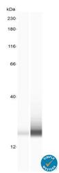
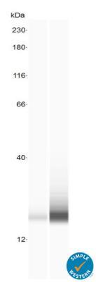
- Simple Western: Lysozyme Antibody [NBP2-61118] - Lane view shows lysates of Ileum stem cells undifferentiated and day 5 differentiated, loaded at 0.2 mg/mL. A specific band was detected for Lysozyme at approximately 20 kDa (as indicated) using 20 ug/mL of Rabbit Anti-Lysozyme Polyclonal Antibody (NBP2-61118). This experiment was conducted under reducing conditions and using the 12-230 kDa separation system.
Supportive validation
- Submitted by
- Novus Biologicals (provider)
- Main image

- Experimental details
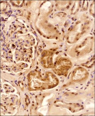
- Immunohistochemistry-Paraffin: Lysozyme Antibody [NBP2-61118] - Analysis of a FFPE tissue section of human kidney with Lysozyme antibody at 1:100 dilution. The staining was developed with HRP-DAB detection method and the counterstaining was performed using hematoxylin. This Lysozyme antibody generated an expected cytoplasmic staining in all the cells with strongest signal in tubular epithelial cells. Some tubules showed more of a punctate staining pattern (vesicular Lysozyme) while all of the tubules depicted a diffused signal (secreted Lysozyme). A subset of cells, especially those from glomeruli, showed nuclear positivity also.
Supportive validation
- Submitted by
- Novus Biologicals (provider)
- Main image
- Experimental details
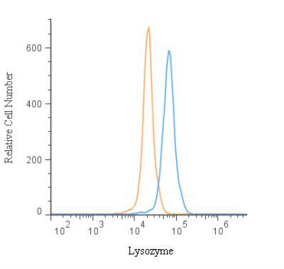
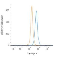
- Flow Cytometry: Lysozyme Antibody [NBP2-61118] - An intracellular stain was performed on THP-1 Cells with NBP2-61118 and a matched isotype control. Cells were fixed with 4% PFA and then permeablized with 0.1% saponin. Cells were incubated in an antibody dilution of 2.5 ug/mL for 30 minutes at room temperature, followed by Rabbit IgG APC-conjugated Secondary Antibody (R&D Systems, F0111).