Explore
 Validate
 Learn
Learn Western blot
 Immunocytochemistry
 Immunohistochemistry
Antibody data
- Antibody Data
- Antigen structure
- References [1]
- Comments [0]
- Validations
- Western blot [1]
- Other assay [1]
Submit
Validation data
Reference
Comment
Report error
- Product number
- PA1-18308 - Provider product page

- Provider
- Invitrogen Antibodies
- Product name
- Anti-Myeloperoxidase Polyclonal Antibody
- Antibody type
- Polyclonal
- Antigen
- Other
- Description
- PA1-18308 detects Myeloperoxidase from human samples. PA1-18308 has been successfully used in immunohistochemistry (paraffin and frozen) and Western blot procedures. The PA1-18308 immunogen is Myeloperoxidase isolated from human polymorphonuclear leucocytes. Reconstitute with 250 µL of distilled water.
- Reactivity
- Human
- Host
- Rabbit
- Isotype
- IgG
- Vial size
- 250 µg
- Concentration
- 1 mg/mL
- Storage
- -20° C, Avoid Freeze/Thaw Cycles
Submitted references Analysis of IL-17(+) cells in facet joints of patients with spondyloarthritis suggests that the innate immune pathway might be of greater relevance than the Th17-mediated adaptive immune response.
Appel H, Maier R, Wu P, Scheer R, Hempfing A, Kayser R, Thiel A, Radbruch A, Loddenkemper C, Sieper J
Arthritis research & therapy 2011 Jun 20;13(3):R95
Arthritis research & therapy 2011 Jun 20;13(3):R95
No comments: Submit comment
Supportive validation
- Submitted by
- Invitrogen Antibodies (provider)
- Main image
- Experimental details
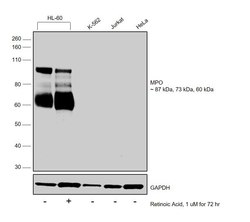
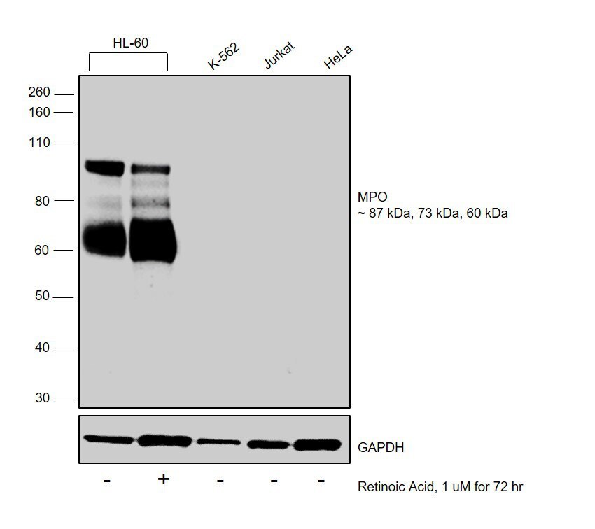
- Western blot was performed using Anti-Myeloperoxidase Polyclonal Antibody (Product # PA1-18308) and 87 kDa, 73 kDa and 60 kDa bands corresponding to MPO was observed across cell lines except K-562, Jurkat and HeLa and was also observed to decrease upon Retinoic Acid treatment. Membrane enriched extracts (30 µg lysate) of HL-60 (Lane 1), HL-60 treated with Retinoic Acid (1uM for 72 Hours) (Lane 2), K-562 (Lane 3), Jurkat (Lane 4) and HeLa (Lane 5) were electrophoresed using NuPAGE™ 4-12% Bis-Tris Protein Gel (Product # NP0322BOX). Resolved proteins were then transferred onto a nitrocellulose membrane (Product # IB23001) by iBlot® 2 Dry Blotting System (Product # IB21001). The blot was probed with the primary antibody (1:1000 dilution) and detected by chemiluminescence with Goat anti-Rabbit IgG (H+L), Superclonal™ Recombinant Secondary Antibody, HRP (Product # A27036, 1:4000 dilution) using the iBright FL 1000 (Product # A32752). Chemiluminescent detection was performed using Novex® ECL Chemiluminescent Substrate Reagent Kit (Product # WP20005).
Supportive validation
- Submitted by
- Invitrogen Antibodies (provider)
- Main image

- Experimental details
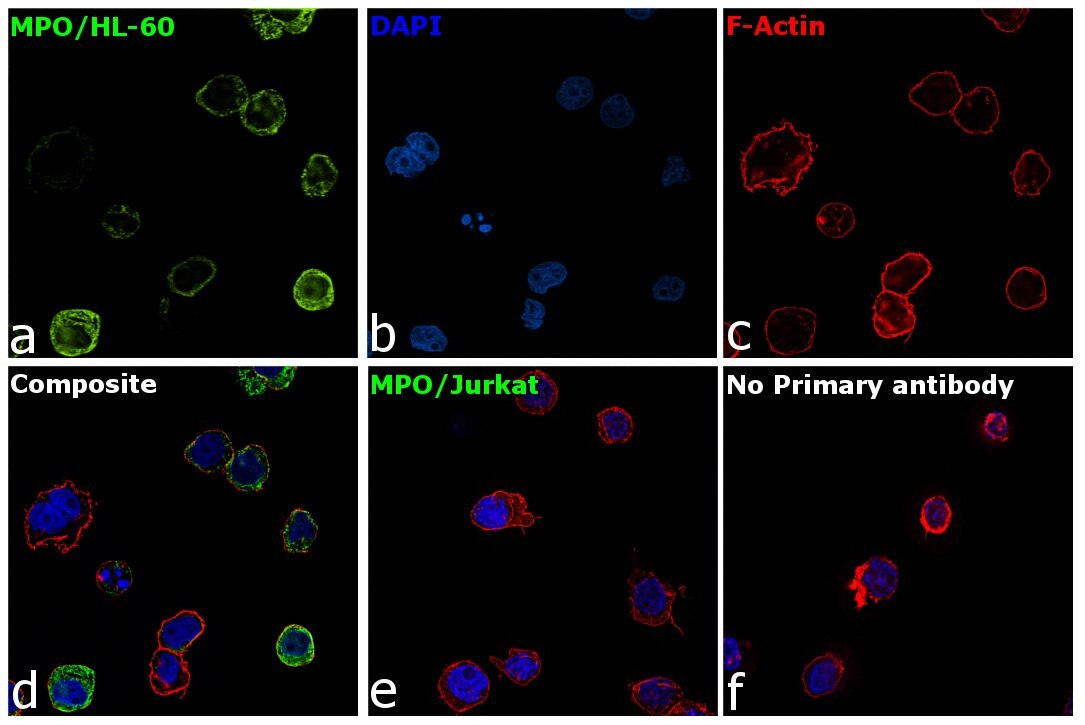
- Immunofluorescence analysis of MPO was performed using HL-60 and Jurkat cells. The cells were fixed with 4% paraformaldehyde for 10 minutes, permeabilized with 0.1% Triton™ X-100 for 15 minutes, and blocked with 2% BSA for 1 hour at room temperature. HL-60 and Jurkat cells were labeled with Myeloperoxidase Polyclonal Antibody (Product # PA1-18308) at 1:100 dilution in 0.1% BSA, incubated at 4 degree Celsius overnight and then labeled with Goat anti-Rabbit IgG (H+L) Superclonal™ Recombinant Secondary Antibody, Alexa Fluor® 488 conjugate (Product # A27034) at a dilution of 1:2000 for 45 minutes at room temperature (Panel a: green). Nuclei (Panel b: blue) were stained with ProLong™ Diamond Antifade Mountant with DAPI (Product # P36962). F-actin (Panel c: red) was stained with Rhodamine Phalloidin (Product # R415). Panel d represents the merged image of HL-60 showing membranous localization. Panel e represents the merged image of Jurkat cells showing no expression for MPO protein. Panel f represents control cells with no primary antibody to assess background. The images were captured at 60X magnification.