Explore
 Validate
 Learn
Learn Western blot
Antibody data
- Antibody Data
- Antigen structure
- References [0]
- Comments [0]
- Validations
- Western blot [2]
- Immunocytochemistry [2]
- Immunohistochemistry [4]
- Flow cytometry [1]
Submit
Validation data
Reference
Comment
Report error
- Product number
- R30233 - Provider product page

- Provider
- NSJ Bioreagents
- Product name
- Myeloperoxidase Antibody / MPO
- Antibody type
- Polyclonal
- Description
- This highly specific Myeloperoxidase antibody is suitable for use in Western blot/Immunohistochemistry/Flow cytometry/Immunofluorescence applications with human, mouse and rat samples.
- Reactivity
- Human, Mouse, Rat
- Host
- Rabbit
- Conjugate
- Unconjugated
- Vial size
- 100 ug
- Concentration
- 0.5mg/ml if reconstituted with 0.2ml sterile DI water
- Storage
- The lyophilized Myeloperoxidase antibody can be stored at 4oC. After reconstitution, aliquot and store at -20oC. Avoid repeated freezing and thawing.
No comments: Submit comment
Supportive validation
- Submitted by
- NSJ Bioreagents (provider)
- Main image
- Experimental details
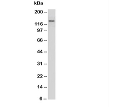
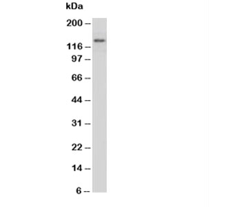
- Western blot testing of Myeloperoxidase antibody and rat brain tissue lysate
- Submitted by
- NSJ Bioreagents (provider)
- Main image
- Experimental details
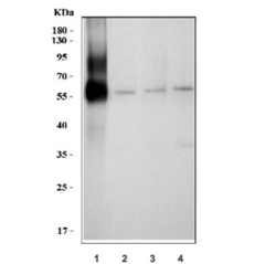
- Western blot testing of 1) human HL60, 2) rat thymus, 3) mouse spleen and 4) mouse thymus tissue with Myeloperoxidase antibody. Expected molecular weight: 59-64 kDa (alpha chain, may be observed at higher molecular weights due to glycosylation), 150+ kDa (glycosylated mature form).

Supportive validation
- Submitted by
- NSJ Bioreagents (provider)
- Main image
- Experimental details
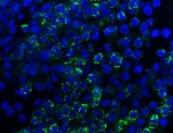
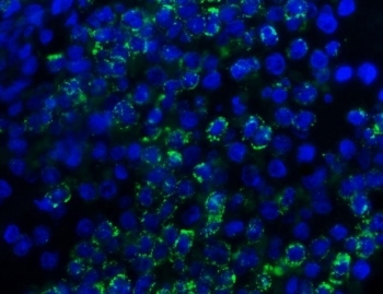
- Immunofluorescent staining of FFPE human spleen tissue with Myeloperoxidase antibody (green) and DAPI nuclear stain (blue). HIER: steam section in pH8 EDTA buffer for 20 min.
- Submitted by
- NSJ Bioreagents (provider)
- Main image

- Experimental details
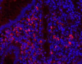
- Immunofluorescent staining of FFPE human appendicitis tissue with Myeloperoxidase antibody (red) and DAPI nuclear stain (blue). HIER: steam section in pH8 EDTA buffer for 20 min.
Supportive validation
- Submitted by
- NSJ Bioreagents (provider)
- Main image
- Experimental details
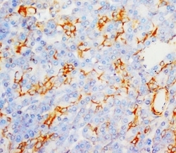
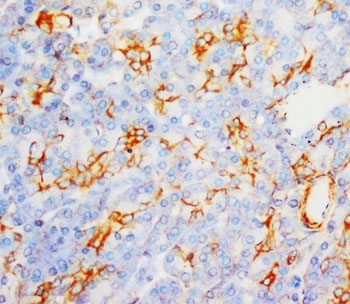
- IHC-P: Myeloperoxidase antibody testing of human liver cancer tissue
- Submitted by
- NSJ Bioreagents (provider)
- Main image
- Experimental details
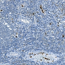
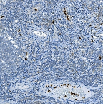
- IHC staining of FFPE rat lymph tissue with Myeloperoxidase antibody. HIER: boil tissue sections in pH8 EDTA for 20 min and allow to cool before testing.
- Submitted by
- NSJ Bioreagents (provider)
- Main image
- Experimental details
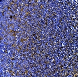
- IHC staining of FFPE human tonsil tissue with Myeloperoxidase antibody. HIER: boil tissue sections in pH8 EDTA for 20 min and allow to cool before testing.
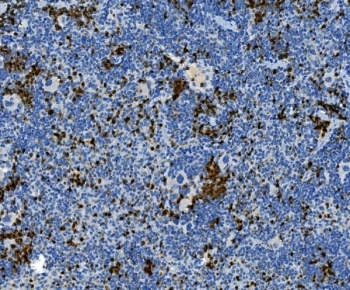
- Submitted by
- NSJ Bioreagents (provider)
- Main image

- Experimental details
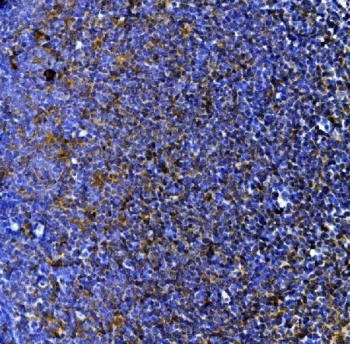
- IHC staining of FFPE mouse spleen tissue with Myeloperoxidase antibody. HIER: boil tissue sections in pH8 EDTA for 20 min and allow to cool before testing.
Supportive validation
- Submitted by
- NSJ Bioreagents (provider)
- Main image
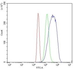
- Experimental details
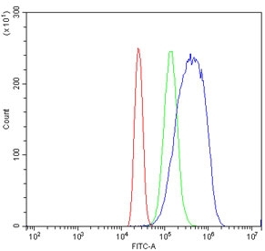
- Flow cytometry testing of human HL60 cells with Myeloperoxidase antibody at 1ug/million cells (blocked with goat sera); Red=cells alone, Green=isotype control, Blue= Myeloperoxidase antibody.