Explore
 Validate
 Learn
Learn Western blot
 Immunocytochemistry
 Immunohistochemistry
Antibody data
- Antibody Data
- Antigen structure
- References [2]
- Comments [0]
- Validations
- Immunocytochemistry [8]
Submit
Validation data
Reference
Comment
Report error
- Product number
- PA1-46203 - Provider product page

- Provider
- Invitrogen Antibodies
- Product name
- NSE Polyclonal Antibody
- Antibody type
- Polyclonal
- Antigen
- Purifed from natural sources
- Description
- Suggested positive control: antigen standard for ENO2 (transient overexpression lysate), rat spinal cord or peripheral nerve homogenate and HEK 293 lysate..
- Reactivity
- Human, Mouse, Rat
- Host
- Rabbit
- Isotype
- IgG
- Vial size
- 100 μL
- Concentration
- Conc. Not Determined
- Storage
- Store at 4°C short term. For long term storage, store at -20°C, avoiding freeze/thaw cycles.
Submitted references The dual anti-inflammatory and antioxidant activities of natural honey promote cell proliferation and neural regeneration in a rat model of colitis.
Valproic acid, a histone deacetylase inhibitor, decreases proliferation of and induces specific neurogenic differentiation of canine adipose tissue-derived stem cells.
Nooh HZ, Nour-Eldien NM
Acta histochemica 2016 Jul;118(6):588-595
Acta histochemica 2016 Jul;118(6):588-595
Valproic acid, a histone deacetylase inhibitor, decreases proliferation of and induces specific neurogenic differentiation of canine adipose tissue-derived stem cells.
Kurihara Y, Suzuki T, Sakaue M, Murayama O, Miyazaki Y, Onuki A, Aoki T, Saito M, Fujii Y, Hisasue M, Tanaka K, Takizawa T
The Journal of veterinary medical science 2014 Jan;76(1):15-23
The Journal of veterinary medical science 2014 Jan;76(1):15-23
No comments: Submit comment
Supportive validation
- Submitted by
- Invitrogen Antibodies (provider)
- Main image
- Experimental details
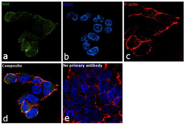
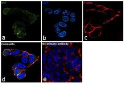
- Immunofluorescence analysis of NSE was performed using 70% confluent log phase HEK-293 cells. The cells were fixed with 4% paraformaldehyde for 10 minutes, permeabilized with 0.1% Triton™ X-100 for 10 minutes, and blocked with 2% BSA for 1 hour at room temperature. The cells were labeled with NSE Polyclonal Antibody (Product # PA1-46203) at 1:100 dilution in 0.1% BSA and incubated overnight at 4 degree and then labeled with Goat anti-Rabbit IgG (H+L) Superclonal™ Secondary Antibody, Alexa Fluor® 488 conjugate (Product # A27034) at a dilution of 1:2000 for 45 minutes at room temperature (Panel a: green). Nuclei (Panel b: blue) were stained with SlowFade® Gold Antifade Mountant with DAPI (Product # S36938). F-actin (Panel c: red) was stained with Rhodamine Phalloidin (Product # R415, 1:300). Panel d represents the merged image showing cytoplasmic and cell membrane localization. Panel e represents control cells with no primary antibody to assess background. The images were captured at 60X magnification.

- Submitted by
- Invitrogen Antibodies (provider)
- Main image
- Experimental details
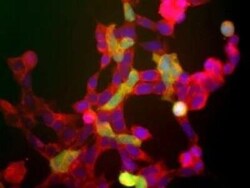
- Immunocytochemistry analysis of NSE in human embryonic kidney cells line 293, which express many neuronal proteins (1). Samples were incubated in NSE polyclonal antibody (Product # PA1-46203). The red channel shows staining which recognizes all of these 293 cells. The green channels shows staining for another neuronal marker to UCHL1. This neuronal gene is apparently activated in a cell density dependent fashion and at this stage only a few cells express this protein. However all cells that express NSE also express UCHL1.

- Submitted by
- Invitrogen Antibodies (provider)
- Main image
- Experimental details
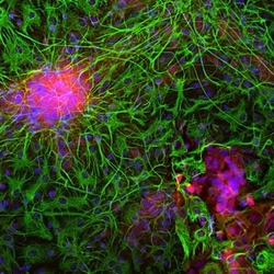
- Immunocytochemistry analysis of NSE in mixed cortical neuron-glial cell culture from E20 rat. Samples were incubated in NSE polyclonal antibody (Product # PA1-46203) using a dilution of 1:500. Neuron specific enolase in red and costained with chicken pAb to GFAP, dilution 1:5,000 in green. The blue is Hoechst staining of nuclear DNA. the NSE antibody labels protein expressed in neuronal cells, while the GFAP antibody stains intermediate filaments in astrocytic and certain other glial cells.

- Submitted by
- Invitrogen Antibodies (provider)
- Main image
- Experimental details
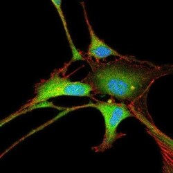
- Immunocytochemistry analysis of NSE in 3T3 cells. Samples were incubated in NSE polyclonal antibody (Product # PA1-46203) followed by Alexa Fluor 488-conjugated Goat to rabbit IgG secondary antibody (green, A). Actin filaments were labeled with Alexa Fluor 568 phalloidin (red, B). DAPI was used to stain the cell nuclei (blue, C).

- Submitted by
- Invitrogen Antibodies (provider)
- Main image

- Experimental details
- Immunocytochemistry analysis of NSE in human embryonic kidney cells line 293, which express many neuronal proteins (1). Samples were incubated in NSE polyclonal antibody (Product # PA1-46203). The red channel shows staining which recognizes all of these 293 cells. The green channels shows staining for another neuronal marker to UCHL1. This neuronal gene is apparently activated in a cell density dependent fashion and at this stage only a few cells express this protein. However all cells that express NSE also express UCHL1.

- Submitted by
- Invitrogen Antibodies (provider)
- Main image

- Experimental details
- Immunocytochemistry analysis of NSE in mixed cortical neuron-glial cell culture from E20 rat. Samples were incubated in NSE polyclonal antibody (Product # PA1-46203) using a dilution of 1:500. Neuron specific enolase in red and costained with chicken pAb to GFAP, dilution 1:5,000 in green. The blue is Hoechst staining of nuclear DNA. the NSE antibody labels protein expressed in neuronal cells, while the GFAP antibody stains intermediate filaments in astrocytic and certain other glial cells.

- Submitted by
- Invitrogen Antibodies (provider)
- Main image

- Experimental details
- Immunocytochemistry analysis of NSE in 3T3 cells. Samples were incubated in NSE polyclonal antibody (Product # PA1-46203) followed by Alexa Fluor 488-conjugated Goat to rabbit IgG secondary antibody (green, A). Actin filaments were labeled with Alexa Fluor 568 phalloidin (red, B). DAPI was used to stain the cell nuclei (blue, C).

- Submitted by
- Invitrogen Antibodies (provider)
- Main image

- Experimental details
- Immunofluorescence analysis of NSE was performed using 70% confluent log phase HEK-293 cells. The cells were fixed with 4% paraformaldehyde for 10 minutes, permeabilized with 0.1% Triton™ X-100 for 10 minutes, and blocked with 2% BSA for 1 hour at room temperature. The cells were labeled with NSE Polyclonal Antibody (Product # PA1-46203) at 1:100 dilution in 0.1% BSA and incubated overnight at 4 degree and then labeled with Goat anti-Rabbit IgG (Heavy Chain) Superclonal™ Secondary Antibody, Alexa Fluor® 488 conjugate (Product # A27034) at a dilution of 1:2000 for 45 minutes at room temperature (Panel a: green). Nuclei (Panel b: blue) were stained with SlowFade® Gold Antifade Mountant with DAPI (Product # S36938). F-actin (Panel c: red) was stained with Rhodamine Phalloidin (Product # R415, 1:300). Panel d represents the merged image showing cytoplasmic and cell membrane localization. Panel e represents control cells with no primary antibody to assess background. The images were captured at 60X magnification.