 Explore
 Validate
 Learn
Learn Western blot
Antibody data
- Antibody Data
- Antigen structure
- References [1]
- Comments [0]
- Validations
- Western blot [3]
- Immunohistochemistry [2]
Submit
Validation data
Reference
Comment
Report error
- Product number
- AF5169 - Provider product page

- Provider
- R&D Systems
- Product name
- Human/Mouse/Rat Enolase 2/Neuron-specific Enolase Antibody
- Antibody type
- Polyclonal
- Description
- Antigen Affinity-purified. Detects human and mouse Enolase 2/Neuron-specific Enolase in direct ELISAs. Detects human, mouse, and rat Enolase 2/Neuron-specific Enolase in Western blots.
- Reactivity
- Human, Mouse, Rat
- Host
- Sheep
- Conjugate
- Unconjugated
- Antigen sequence
P09104- Isotype
- IgG
- Vial size
- 100 ug
- Concentration
- LYOPH
- Storage
- Use a manual defrost freezer and avoid repeated freeze-thaw cycles. 12 months from date of receipt, -20 to -70 °C as supplied. 1 month, 2 to 8 °C under sterile conditions after reconstitution. 6 months, -20 to -70 °C under sterile conditions after reconstitution.
Submitted references Inflammation-induced reversible switch of the neuron-specific enolase promoter from Purkinje neurons to Bergmann glia.
Sawada Y, Konno A, Nagaoka J, Hirai H
Scientific reports 2016 Jun 13;6:27758
Scientific reports 2016 Jun 13;6:27758
No comments: Submit comment
Supportive validation
- Submitted by
- R&D Systems (provider)
- Main image
- Experimental details
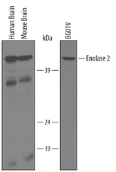
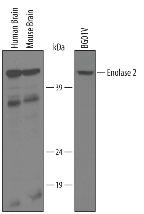
- Detection of Human/Mouse Enolase 2 by Western Blot. Western blot shows lysates of human brain, mouse brain tissue, and BG01V human embryonic stem cells. PVDF membrane was probed with 1 µg/mL of Human/Mouse Enolase 2 Antigen Affinity-purified Polyclonal Antibody (Catalog # AF5169) followed by HRP-conjugated Anti-Sheep IgG Secondary Antibody (Catalog # HAF016). A specific band was detected for Enolase 2 at approximately 46 kDa (as indicated). This experiment was conducted under reducing conditions and using Immunoblot Buffer Group 8.
- Submitted by
- R&D Systems (provider)
- Main image
- Experimental details
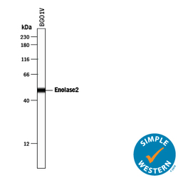
- Detection of Human Enolase 2/Neuron-specific Enolase by Simple WesternTM. Simple Western lane view shows lysates of BG01V human embryonic stem cells, loaded at 0.2 mg/mL. A specific band was detected for Enolase 2/Neuron-specific Enolase at approximately 50 kDa (as indicated) using 10 µg/mL of Sheep Anti-Human/Mouse Enolase 2/Neuron-specific Enolase Antigen Affinity-purified Polyclonal Antibody (Catalog # AF5169) followed by 1:50 dilution of HRP-conjugated Anti-Sheep IgG Secondary Antibody (Catalog # HAF016). This experiment was conducted under reducing conditions and using the 12-230 kDa separation system.

- Submitted by
- R&D Systems (provider)
- Main image
- Experimental details
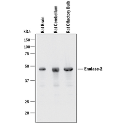
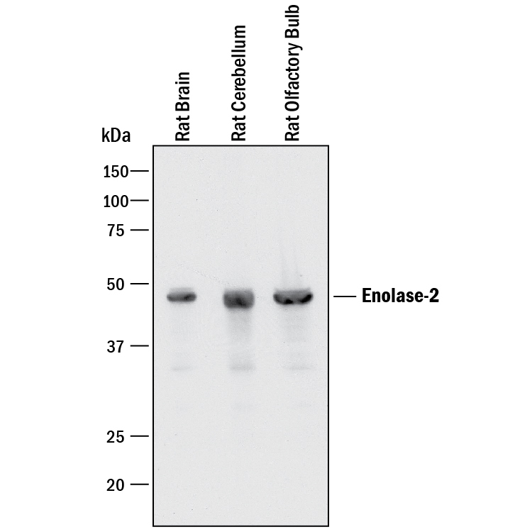
- Detection of Rat Enolase 2/Neuron-specific Enolase by Western Blot. Western blot shows lysates of rat brain tissue, rat cerebellum tissue, and rat olfactroy bulb tissue. PVDF membrane was probed with 0.2 µg/mL of Sheep Anti-Human/Mouse Enolase 2/Neuron-specific Enolase Antigen Affinity-purified Polyclonal Antibody (Catalog # AF5169) followed by HRP-conjugated Anti-Sheep IgG Secondary Antibody (Catalog # HAF016). A specific band was detected for Enolase 2/Neuron-specific Enolase at approximately 47 kDa (as indicated). This experiment was conducted under reducing conditions and using Immunoblot Buffer Group 1.
Supportive validation
- Submitted by
- R&D Systems (provider)
- Main image

- Experimental details
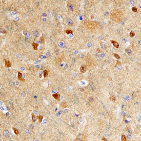
- Enolase 2 in Human Brain. Enolase 2 was detected in immersion fixed paraffin-embedded sections of human brain (cortex) using Human/Mouse Enolase 2 Antigen Affinity-purified Polyclonal Antibody (Catalog # AF5169) at 10 µg/mL overnight at 4 °C. Before incubation with the primary antibody, tissue was subjected to heat-induced epitope retrieval using Antigen Retrieval Reagent-Basic (Catalog # CTS013). Tissue was stained using the Anti-Sheep HRP-DAB Cell & Tissue Staining Kit (brown; Catalog # CTS019) and counterstained with hematoxylin (blue). Specific staining was localized to cytoplasm. View our protocol for Chromogenic IHC Staining of Paraffin-embedded Tissue Sections.
- Submitted by
- R&D Systems (provider)
- Main image
- Experimental details
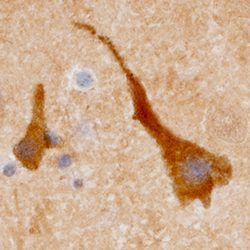
- Enolase 2 in Human Brain. Enolase 2 was detected in immersion fixed paraffin-embedded sections of human brain (cortex) using Human/Mouse Enolase 2 Antigen Affinity-purified Polyclonal Antibody (Catalog # AF5169) at 10 µg/mL overnight at 4 °C. Before incubation with the primary antibody, tissue was subjected to heat-induced epitope retrieval using Antigen Retrieval Reagent-Basic (Catalog # CTS013). Tissue was stained using the Anti-Sheep HRP-DAB Cell & Tissue Staining Kit (brown; Catalog # CTS019) and counterstained with hematoxylin (blue). Specific staining was localized to cytoplasm. View our protocol for Chromogenic IHC Staining of Paraffin-embedded Tissue Sections.
