Explore
 Validate
 Learn
Learn Western blot
 Immunoprecipitation
Antibody data
- Antibody Data
- Antigen structure
- References [6]
- Comments [0]
- Validations
- Western blot [2]
- Immunohistochemistry [1]
Submit
Validation data
Reference
Comment
Report error
- Product number
- AF934 - Provider product page

- Provider
- R&D Systems
- Product name
- Human/Mouse/Rat Cathepsin X/Z/P Antibody
- Antibody type
- Polyclonal
- Description
- Antigen Affinity-purified. Detects human Cathepsin X/Z/P in direct ELISAs and Western blots. In direct ELISAs, less than 5% cross-reactivity with recombinant human Cathepsin C is observed.
- Reactivity
- Human, Mouse, Rat
- Host
- Goat
- Conjugate
- Unconjugated
- Antigen sequence
Q9UBR2- Isotype
- IgG
- Vial size
- 100 ug
- Concentration
- LYOPH
- Storage
- Use a manual defrost freezer and avoid repeated freeze-thaw cycles. 12 months from date of receipt, -20 to -70 °C as supplied. 1 month, 2 to 8 °C under sterile conditions after reconstitution. 6 months, -20 to -70 °C under sterile conditions after reconstitution.
Submitted references Cysteine cathepsins: Their biological and molecular significance in cancer stem cells.
Increased expression and altered localization of cathepsin Z are associated with progression to jaundice stage in primary biliary cholangitis.
Cathepsin X Cleaves Profilin 1 C-Terminal Tyr139 and Influences Clathrin-Mediated Endocytosis.
Distinct functions of macrophage-derived and cancer cell-derived cathepsin Z combine to promote tumor malignancy via interactions with the extracellular matrix.
Prognostic and predictive value of cathepsin X in serum from colorectal cancer patients.
Three-dimensional invasion of macrophages is mediated by cysteine cathepsins in protrusive podosomes.
Pišlar A, Jewett A, Kos J
Seminars in cancer biology 2018 Dec;53:168-177
Seminars in cancer biology 2018 Dec;53:168-177
Increased expression and altered localization of cathepsin Z are associated with progression to jaundice stage in primary biliary cholangitis.
Aiba Y, Harada K, Ito M, Suematsu T, Aishima S, Hitomi Y, Nishida N, Kawashima M, Takatsuki M, Eguchi S, Shimoda S, Nakamura H, Komori A, Abiru S, Nagaoka S, Migita K, Yatsuhashi H, Tokunaga K, Nakamura M
Scientific reports 2018 Aug 7;8(1):11808
Scientific reports 2018 Aug 7;8(1):11808
Cathepsin X Cleaves Profilin 1 C-Terminal Tyr139 and Influences Clathrin-Mediated Endocytosis.
Pečar Fonović U, Kos J
PloS one 2015;10(9):e0137217
PloS one 2015;10(9):e0137217
Distinct functions of macrophage-derived and cancer cell-derived cathepsin Z combine to promote tumor malignancy via interactions with the extracellular matrix.
Akkari L, Gocheva V, Kester JC, Hunter KE, Quick ML, Sevenich L, Wang HW, Peters C, Tang LH, Klimstra DS, Reinheckel T, Joyce JA
Genes & development 2014 Oct 1;28(19):2134-50
Genes & development 2014 Oct 1;28(19):2134-50
Prognostic and predictive value of cathepsin X in serum from colorectal cancer patients.
Vižin T, Christensen IJ, Wilhelmsen M, Nielsen HJ, Kos J
BMC cancer 2014 Apr 13;14:259
BMC cancer 2014 Apr 13;14:259
Three-dimensional invasion of macrophages is mediated by cysteine cathepsins in protrusive podosomes.
Jevnikar Z, Mirković B, Fonović UP, Zidar N, Švajger U, Kos J
European journal of immunology 2012 Dec;42(12):3429-41
European journal of immunology 2012 Dec;42(12):3429-41
No comments: Submit comment
Supportive validation
- Submitted by
- R&D Systems (provider)
- Main image

- Experimental details
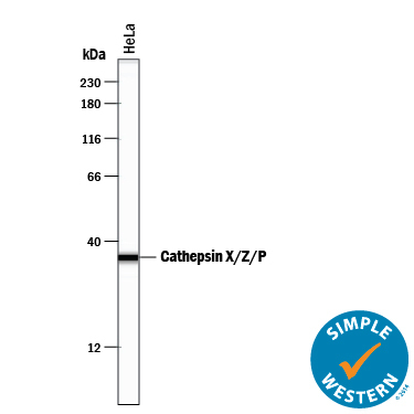
- Detection of Human Cathepsin X/Z/P by Simple Western<SUP abp="263">TM. Simple Western lane view shows lysates of HeLa human cervical epithelial carcinoma cell line, loaded at 0.2 mg/mL. A specific band was detected for Cathepsin X/Z/P at approximately 36 kDa (as indicated) using 5 µg/mL of Goat Anti-Human/Mouse/Rat Cathepsin X/Z/P Antigen Affinity-purified Polyclonal Antibody (Catalog # AF934) followed by 1:50 dilution of HRP-conjugated Anti-Goat IgG Secondary Antibody (Catalog # HAF109). This experiment was conducted under reducing conditions and using the 12-230 kDa separation system.
- Submitted by
- R&D Systems (provider)
- Main image
- Experimental details
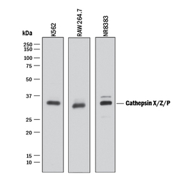
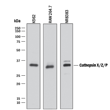
- Detection of Human, Mouse, and Rat Cathepsin X/Z/P by Western Blot. Western blot shows lysates of K562 human chronic myelogenous leukemia cell line, RAW 264.7 mouse monocyte/macrophage cell line, and NR8383 rat alveolar macrophage cell line. PVDF membrane was probed with 1 µg/mL of Goat Anti-Human/ Mouse/Rat Cathepsin X/Z/P Antigen Affinity-purified Polyclonal Antibody (Catalog # AF934) followed by HRP-conjugated Anti-Goat IgG Secondary Antibody (Catalog # HAF017). A specific band was detected for Cathepsin X/Z/P at approximately 34 kDa (as indicated). This experiment was conducted under reducing conditions and using Immunoblot Buffer Group 1.
Supportive validation
- Submitted by
- R&D Systems (provider)
- Main image

- Experimental details
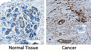
- Cathepsin X/Z/P in Human Breast and Breast Cancer Tissue. Cathepsin X/Z/P was detected in immersion fixed paraffin-embedded sections of normal human breast and breast cancer tissue using Goat Anti-Human/Mouse/Rat Cathepsin X/Z/P Antigen Affinity-purified Polyclonal Antibody (Catalog # AF934) at 3 µg/mL overnight at 4 °C. Tissue was stained using the Anti-Goat HRP-DAB Cell & Tissue Staining Kit (brown; Catalog # CTS008) and counterstained with hematoxylin (blue). Specific staining was localized to stromal and endothelial cells. View our protocol for Chromogenic IHC Staining of Paraffin-embedded Tissue Sections.