 Explore
 Validate
 Learn
Learn Western blot
 ELISA
ELISA Immunocytochemistry
Antibody data
- Antibody Data
- Antigen structure
- References [3]
- Comments [0]
- Validations
- Immunocytochemistry [2]
- Immunohistochemistry [2]
- Flow cytometry [2]
- Other assay [2]
Submit
Validation data
Reference
Comment
Report error
- Product number
- MA5-17182 - Provider product page

- Provider
- Invitrogen Antibodies
- Product name
- Somatostatin Monoclonal Antibody (7G5)
- Antibody type
- Monoclonal
- Antigen
- Purifed from natural sources
- Description
- MA5-17182 targets SST in FACS, IHC, indirect ELISA, and WB applications and shows reactivity with Human samples. The MA5-17182 immunogen is purified recombinant fragment of human SST (amino acids: 1-116) expressed in E. Coli. MA5-17182 detects SST which has a predicted molecular weight of approximately 12.7kDa.
- Reactivity
- Human
- Host
- Mouse
- Isotype
- IgG
- Antibody clone number
- 7G5
- Vial size
- 100 μg
- Concentration
- 1 mg/mL
- Storage
- Store at 4°C short term. For long term storage, store at -20°C, avoiding freeze/thaw cycles.
Submitted references Human iPSC Modeling of Genetic Febrile Seizure Reveals Aberrant Molecular and Physiological Features Underlying an Impaired Neuronal Activity.
Transcriptomic and epigenomic dynamics associated with development of human iPSC-derived GABAergic interneurons.
Flavonoid Glycosides of Polygonum capitatum Protect against Inflammation Associated with Helicobacter pylori Infection.
Scalise S, Zannino C, Lucchino V, Lo Conte M, Scaramuzzino L, Cifelli P, D'Andrea T, Martinello K, Fucile S, Palma E, Gambardella A, Ruffolo G, Cuda G, Parrotta EI
Biomedicines 2022 May 5;10(5)
Biomedicines 2022 May 5;10(5)
Transcriptomic and epigenomic dynamics associated with development of human iPSC-derived GABAergic interneurons.
Inglis GAS, Zhou Y, Patterson DG, Scharer CD, Han Y, Boss JM, Wen Z, Escayg A
Human molecular genetics 2020 Aug 29;29(15):2579-2595
Human molecular genetics 2020 Aug 29;29(15):2579-2595
Flavonoid Glycosides of Polygonum capitatum Protect against Inflammation Associated with Helicobacter pylori Infection.
Zhang S, Mo F, Luo Z, Huang J, Sun C, Zhang R
PloS one 2015;10(5):e0126584
PloS one 2015;10(5):e0126584
No comments: Submit comment
Supportive validation
- Submitted by
- Invitrogen Antibodies (provider)
- Main image

- Experimental details
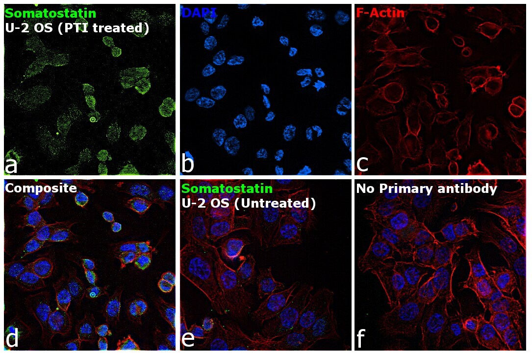
- Immunofluorescence analysis of Somatostatin was performed using 70% confluent log phase U-2 OS cells. The cells were fixed with 4% paraformaldehyde for 10 minutes, permeabilized with 0.1% Triton™ X-100 for 15 minutes, and blocked with 2% BSA for 45 minutes at room temperature. The cells were labeled with Somatostatin Monoclonal Antibody (7G5) (Product # MA5-17182) at 1:200 dilution in 0.1% BSA, incubated at 4 degree celsius overnight and then labeled with Goat anti-Mouse IgG (H+L) Highly Cross-Adsorbed Secondary Antibody, Alexa Fluor Plus 488 (Product # A32723), (1:3000 dilution), for 45 minutes at room temperature (Panel a: Green). Nuclei (Panel b: Blue) were stained with ProLong™ Diamond Antifade Mountant with DAPI (Product # P36962). F-actin (Panel c: Red) was stained with Rhodamine Phalloidin (Product # R415, 1:300 dilution). Panel d represents the merged image showing cytoplasmic localization. Panel e represents merged image for untreated U-2 OS cells showing no staining for Somatostatin. Panel f represents control cells with no primary antibody to assess background. The images were captured at 60X magnification.
- Submitted by
- Invitrogen Antibodies (provider)
- Main image
- Experimental details
- Immunofluorescence analysis of Somatostatin was performed using 70% confluent log phase U-2 OS cells. The cells were fixed with 4% paraformaldehyde for 10 minutes, permeabilized with 0.1% Triton™ X-100 for 15 minutes, and blocked with 2% BSA for 45 minutes at room temperature. The cells were labeled with Somatostatin Monoclonal Antibody (7G5) (Product # MA5-17182) at 1:200 dilution in 0.1% BSA, incubated at 4 degree celsius overnight and then labeled with Goat anti-Mouse IgG (H+L) Highly Cross-Adsorbed Secondary Antibody, Alexa Fluor Plus 488 (Product # A32723), (1:3000 dilution), for 45 minutes at room temperature (Panel a: Green). Nuclei (Panel b: Blue) were stained with ProLong™ Diamond Antifade Mountant with DAPI (Product # P36962). F-actin (Panel c: Red) was stained with Rhodamine Phalloidin (Product # R415, 1:300 dilution). Panel d represents the merged image showing cytoplasmic localization. Panel e represents merged image for untreated U-2 OS cells showing no staining for Somatostatin. Panel f represents control cells with no primary antibody to assess background. The images were captured at 60X magnification.

Supportive validation
- Submitted by
- Invitrogen Antibodies (provider)
- Main image
- Experimental details
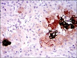
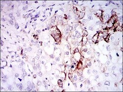
- Immunohistochemical analysis of paraffin-embedded pancreas tissues using SST monoclonal antibody (Product # MA5-17182) followed with DAB staining.
- Submitted by
- Invitrogen Antibodies (provider)
- Main image
- Experimental details
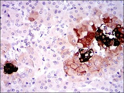
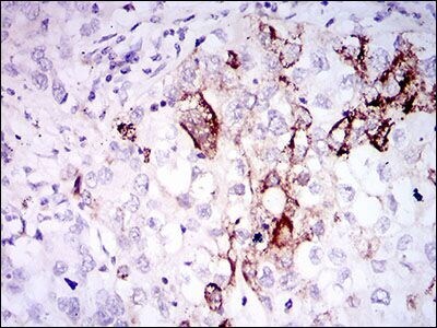
- Immunohistochemical analysis of paraffin-embedded lung cancer tissues using SST monoclonal antibody (Product # MA5-17182) followed with DAB staining.
Supportive validation
- Submitted by
- Invitrogen Antibodies (provider)
- Main image

- Experimental details
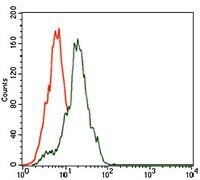
- Flow cytometric analysis of HepG2 cells using SST monoclonal antibody (Product # MA5-17182) (green) and negative control (red).
- Submitted by
- Invitrogen Antibodies (provider)
- Main image

- Experimental details
- Flow cytometric analysis of HepG2 cells using SST monoclonal antibody (Product # MA5-17182) (green) and negative control (red).

Supportive validation
- Submitted by
- Invitrogen Antibodies (provider)
- Main image
- Experimental details
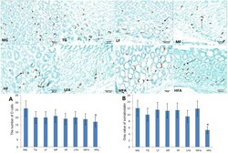
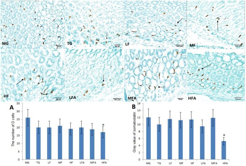
- Fig 6 The staining of D cells of Gastric mucosa-associated lymphoid tissue (MALT) lymphoma (PVx200). Normal mice (without H . pylori infection) were assigned as a control group (CG group) and mice models infected with H . pylori were divided into model group (MG group, only treated with saline solution), triple combination therapy group (TG group, the daily medicine intake is 0.5 ug clarithromycin, 0.02 ug omeprazole, and 1 ug amoxicillin), low/middle/high concentrations of flavonoid glycoside group (LF/MF/HF group, treated with one daily dose of flavonoid glycoside at 32/64/128 ug), low/middle/high concentrations of flavonoid glycoside and common concentration of amoxicillin group (LFA/MFA/HFA group, treated with one daily dose of Flavonoid glycoside at 32/64/128 ug and amoxicillin at 1 ug). Analysis of G cells and somatostatin gray value in each group (n = 10). * P < 0.05 vs model group.
- Submitted by
- Invitrogen Antibodies (provider)
- Main image
- Experimental details
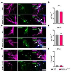
- Types of interneurons generated from iPSCs. Immunofluorescence analysis of idNs stained with antibodies against interneuronal subtypes markers ( A ) somatostatin (SST), ( B ) calretinin (CALB2), and ( C ) calbindin (CALB1). In each group of images, WT cells are shown in the upper panel, while SCN1A M145T idNs are shown in the lower panel. White arrows in the merged images indicate neurons expressing the interneuronal makers indicated (63x magnification). ( D - F ) Quantification of percentage of MAP2 + neurons co-expressing interneuronal markers immunostained in panels ( A - C ). About 9-1% of idNs express SST and CALB2, while CALB1 is present in less than 1% percent of neurons. At least 200 cells were counted for each bar, and data are presented as mean +- SEM of two independent experiments.
