Explore
 Validate
 Learn
Learn Western blot
Antibody data
- Antibody Data
- Antigen structure
- References [0]
- Comments [0]
- Validations
- Western blot [3]
- Immunocytochemistry [2]
- Immunohistochemistry [3]
Submit
Validation data
Reference
Comment
Report error
- Product number
- PA5-18726 - Provider product page

- Provider
- Invitrogen Antibodies
- Product name
- G6PD Polyclonal Antibody
- Antibody type
- Polyclonal
- Antigen
- Synthetic peptide
- Description
- This antibody is predicted to react with canine, mouse and rat based on sequence homology. This antibody is tested in Peptide ELISA: antibody detection limit dilution 8,000.
- Reactivity
- Human
- Host
- Goat
- Isotype
- IgG
- Vial size
- 100 μg
- Concentration
- 0.5 mg/mL
- Storage
- -20°C, Avoid Freeze/Thaw Cycles
No comments: Submit comment
Supportive validation
- Submitted by
- Invitrogen Antibodies (provider)
- Main image

- Experimental details
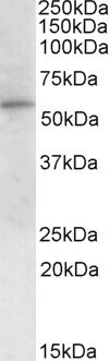
- Western blot analysis of G6PD using G6PD Polyclonal Antibody (Product # PA5-18726) (1 µg/mL) in staining of Human Peripheral Blood Mononucleocytes lysate (35 µg protein in RIPA buffer). Primary incubation was 1 hour. Detected by chemiluminescence.
- Submitted by
- Invitrogen Antibodies (provider)
- Main image
- Experimental details
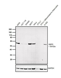
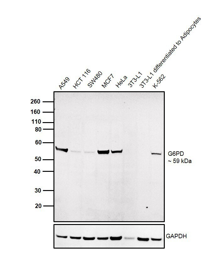
- Western blot was performed using Anti-G6PD Goat Polyclonal Antibody (Product # PA5-18726) and a 59 kDa band corresponding to G6PD was observed across cell lines tested. Whole cell extracts (30 µg lysate) of A549 (Lane 1), HCT 116 (Lane 2), SW480 (Lane 2), MCF7 (Lane 4), HeLa (Lane 5), 3T3-L1 (Lane 6), 3T3-L1 differentiated to Adipocytes (Lane 7) and K-562 (Lane 8) were electrophoresed using NuPAGE™ 4-12% Bis-Tris Protein Gel (Product # NP0322BOX). Resolved proteins were then transferred onto a nitrocellulose membrane (Product # IB23001) by iBlot® 2 Dry Blotting System (Product # IB21001). The blot was probed with the primary antibody (1:500 dilution) and detected by chemiluminescence with Rabbit anti-Goat IgG Heavy Chain, Superclonal™ Recombinant Secondary Antibody, HRP (Product # A27014, 1:4000 dilution) using the iBright FL 1000 (Product # A32752). Chemiluminescent detection was performed using Novex® ECL Chemiluminescent Substrate Reagent Kit (Product # WP20005). This product does not show reactivity to mouse G6PD, as no band was observed in mouse 3T3-L1 cell line when differentiated to adipocytes.
- Submitted by
- Invitrogen Antibodies (provider)
- Main image
- Experimental details
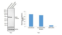
- Knockdown of G6PD was achieved by transfecting HeLa cells with G6PD specific siRNAs (Silencer® select Product # s5447). Western blot analysis (Fig. a) was performed whole cell extracts from the HeLa knockdown cells (lane 3), non-specific scrambled siRNA transfected cells (lane 2) and untransfected cells (lane 1). The blots were probed with G6PD Polyclonal Antibody (Product # PA5-18726, 1:500) and Rabbit anti-Goat IgG Heavy Chain Superclonal™ Recombinant Secondary Antibody, HRP (Product # A27014, 1:4000 dilution). Densitometric analysis of this western blot is shown in histogram (Fig. b). Decrease in signal upon siRNA mediated knock down confirms that antibody is specific to G6PD.

Supportive validation
- Submitted by
- Invitrogen Antibodies (provider)
- Main image

- Experimental details
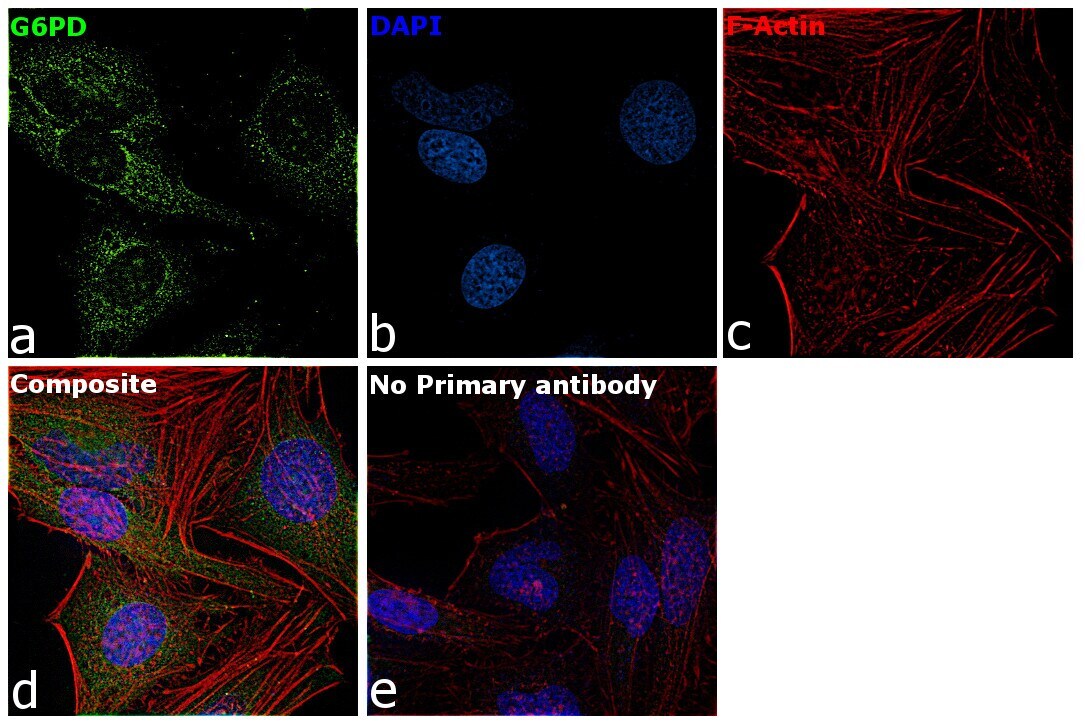
- Immunofluorescence analysis of G6PD was performed using HeLa cells. The cells were fixed with 4% paraformaldehyde for 10 minutes, permeabilized with 0.1% Triton™ X-100 for 15 minutes, and blocked with 2% BSA for 1 hour at room temperature. The cells were labeled with G6PD Goat Polyclonal Antibody (Product # PA5-18726) at 5 µg/mL in 0.1% BSA and incubated overnight at 4 degree and then labeled with Rabbit anti-Goat IgG (H+L) Superclonal™ Recombinant Secondary Antibody, Alexa Fluor® 488 conjugate (Product # A27012) at a dilution of 1:2000 for 45 minutes at room temperature (Panel a: green). Nuclei (Panel b: blue) were stained with ProLong™ Diamond Antifade Mountant with DAPI (Product # P36962). F-actin (Panel c: red) was stained with Rhodamine Phalloidin (Product # R415, 1:300). Panel d represents the composite image showing Nuclear and Cytoplasmic localization of G6PD. Panel e represents control cells with no primary antibody to assess background. The images were captured at 60X magnification.

- Submitted by
- Invitrogen Antibodies (provider)
- Main image

- Experimental details
- Immunofluorescence analysis of G6PD was performed using HeLa cells. The cells were fixed with 4% paraformaldehyde for 10 minutes, permeabilized with 0.1% Triton™ X-100 for 15 minutes, and blocked with 2% BSA for 1 hour at room temperature. The cells were labeled with G6PD Goat Polyclonal Antibody (Product # PA5-18726) at 5 µg/mL in 0.1% BSA and incubated overnight at 4 degree and then labeled with Rabbit anti-Goat IgG Heavy Chain Superclonal™ Recombinant Secondary Antibody, Alexa Fluor® 488 conjugate (Product # A27012) at a dilution of 1:2000 for 45 minutes at room temperature (Panel a: green). Nuclei (Panel b: blue) were stained with ProLong™ Diamond Antifade Mountant with DAPI (Product # P36962). F-actin (Panel c: red) was stained with Rhodamine Phalloidin (Product # R415, 1:300). Panel d represents the composite image showing Nuclear and Cytoplasmic localization of G6PD. Panel e represents control cells with no primary antibody to assess background. The images were captured at 60X magnification.
Supportive validation
- Submitted by
- Invitrogen Antibodies (provider)
- Main image
- Experimental details
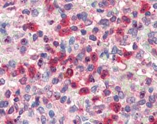
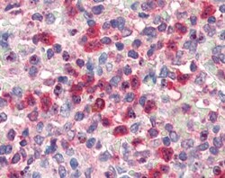
- Immunohistochemistry analysis of G6PD in human spleen. Samples were incubated with G6PD polyclonal antibody (Product # PA5-18726) using a dilution of 5 µg/mL. Formalin-fixed, paraffin-embedded tissue after heat-induced antigen retrieval.

- Submitted by
- Invitrogen Antibodies (provider)
- Main image
- Experimental details
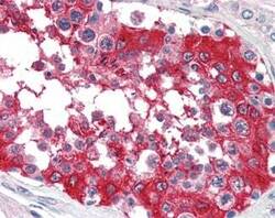
- Immunohistochemistry analysis of G6PD in human testis. Samples were incubated with G6PD polyclonal antibody (Product # PA5-18726) using a dilution of 5 µg/mL. Formalin-fixed, paraffin-embedded tissue after heat-induced antigen retrieval.

- Submitted by
- Invitrogen Antibodies (provider)
- Main image

- Experimental details
- Immunohistochemistry analysis of G6PD in human spleen. Samples were incubated with G6PD polyclonal antibody (Product # PA5-18726) using a dilution of 5 µg/mL. Formalin-fixed, paraffin-embedded tissue after heat-induced antigen retrieval.