Explore
 Validate
 Learn
Learn Western blot
 Immunocytochemistry
Antibody data
- Antibody Data
- Antigen structure
- References [2]
- Comments [0]
- Validations
- Immunocytochemistry [1]
- Immunohistochemistry [1]
- Other assay [1]
Submit
Validation data
Reference
Comment
Report error
- Product number
- PA5-27359 - Provider product page

- Provider
- Invitrogen Antibodies
- Product name
- G6PD Polyclonal Antibody
- Antibody type
- Polyclonal
- Antigen
- Recombinant full-length protein
- Description
- Recommended positive controls: 293T, A431, H1299, HeLaS3, HepG2, Raji, Neuro 2A, C8D30, NIH-3T3, Raw264.7, C2C12, PC-12, Rat2. Predicted reactivity: Rat (93%), Xenopus laevis (82%), Rabbit (95%), Sheep (92%), Rhesus Monkey (99%), Bovine (95%). Store product as a concentrated solution. Centrifuge briefly prior to opening the vial.
- Reactivity
- Human, Mouse, Rat
- Host
- Rabbit
- Isotype
- IgG
- Vial size
- 100 μL
- Concentration
- 0.81 mg/mL
- Storage
- Store at 4°C short term. For long term storage, store at -20°C, avoiding freeze/thaw cycles.
Submitted references Breast cancer stem cells rely on fermentative glycolysis and are sensitive to 2-deoxyglucose treatment.
p63 isoforms regulate metabolism of cancer stem cells.
Ciavardelli D, Rossi C, Barcaroli D, Volpe S, Consalvo A, Zucchelli M, De Cola A, Scavo E, Carollo R, D'Agostino D, Forlì F, D'Aguanno S, Todaro M, Stassi G, Di Ilio C, De Laurenzi V, Urbani A
Cell death & disease 2014 Jul 17;5(7):e1336
Cell death & disease 2014 Jul 17;5(7):e1336
p63 isoforms regulate metabolism of cancer stem cells.
D'Aguanno S, Barcaroli D, Rossi C, Zucchelli M, Ciavardelli D, Cortese C, De Cola A, Volpe S, D'Agostino D, Todaro M, Stassi G, Di Ilio C, Urbani A, De Laurenzi V
Journal of proteome research 2014 Apr 4;13(4):2120-36
Journal of proteome research 2014 Apr 4;13(4):2120-36
No comments: Submit comment
Supportive validation
- Submitted by
- Invitrogen Antibodies (provider)
- Main image
- Experimental details
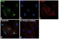
- Immunofluorescence analysis of G6PD was performed using HeLa cells. The cells were fixed with 4% paraformaldehyde for 10 minutes, permeabilized with 0.1% Triton™ X-100 for 15 minutes, and blocked with 2% BSA for 1 hour at room temperature. The cells were labeled with G6PD Goat Polyclonal Antibody (Product # PA5-27359) at 1:100 dilution in 0.1% BSA and incubated overnight at 4 degree and then labeled with Goat anti-Rabbit IgG (Heavy Chain) Superclonal™ Recombinant Secondary Antibody, Alexa Fluor® 488 conjugate (Product # A27034) at a dilution of 1:2000 for 45 minutes at room temperature (Panel a: green). Nuclei (Panel b: blue) were stained with ProLong™ Diamond Antifade Mountant with DAPI (Product # P36962). F-actin (Panel c: red) was stained with Rhodamine Phalloidin (Product # R415, 1:300). Panel d represents the composite image showing Nuclear and Cytoplasmic localization of G6PD. Panel e represents control cells with no primary antibody to assess background. The images were captured at 60X magnification.

Supportive validation
- Submitted by
- Invitrogen Antibodies (provider)
- Main image
- Experimental details
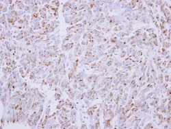
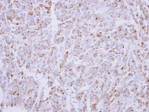
- Immunohistochemical analysis of paraffin-embedded U87 xenograft, using G6PD (Product # PA5-27359) antibody at 1:300 dilution. Antigen Retrieval: EDTA based buffer, pH 8.0, 15 min.
Supportive validation
- Submitted by
- Invitrogen Antibodies (provider)
- Main image

- Experimental details
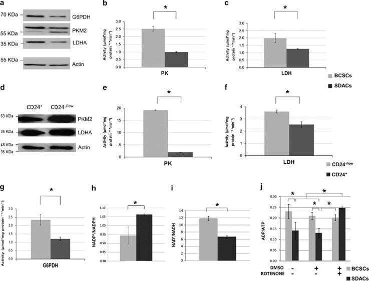
- Figure 4 The expression levels and activities of glycolytic enzymes are increased in BCSCs and correlate with altered energy and redox status of BCSCs. ( a ) WB showing PKM2, LDH-A, and G6PDH expression in BCSCs compared with SDACs. beta -Actin is used as a protein-loading control. A representative WB of three independent experiments is shown. ( b ) PK and ( c ) LDH, and enzyme activities evaluated in spheres (BCSCs) as compared with differentiated cells (SDACs). Bar graphs show the mean activity ( n =6) expressed as mu mol/mg Protein /min, and bars represent the S.E.M. ( d ) WB showing the expression of PKM2 and LDH-A in cells sorted for CD24 expression. beta -Actin is used as a loading control. ( e ) PK and ( f ) LDH activity in CD24 -/low cells compared with CD24 + BCSCs. Bar graphs show the mean activity ( n =6) expressed as mu mol/mg Protein /min, and bars represent the S.E.M. ( g ) G6PDH enzyme activity evaluated in spheres (BCSCs) as compared with differentiated cells (SDACs). Bar graphs show the mean activity ( n =6) expressed as mu mol/mg Protein /min, and bars represent the S.E.M. ( h ) NADP + /NADPH, ( i ) NAD + /NADH, and ( j ) ADP/ATP ratios in BCSCs compared with SDACs. Treatment with rotenone does not affect ATP production in BCSCs, whereas strongly increases the ADP/ATP ratio and inhibits mitochondrial ATP production in SDACs. No changes in the ADP/ATP ratio are observed in BCSCs and SDACs after addition of dimethylsulphoxide (DMSO) alone used as a vehicle con