Explore
 Validate
 Learn
Learn Western blot
 ELISA
ELISAAntibody data
- Antibody Data
- Antigen structure
- References [0]
- Comments [0]
- Validations
- Western blot [3]
- Flow cytometry [2]
Submit
Validation data
Reference
Comment
Report error
- Product number
- NBP2-37822 - Provider product page

- Provider
- Novus Biologicals
- Product name
- Mouse Monoclonal c-Myc Antibody
- Antibody type
- Monoclonal
- Description
- Protein A purified. Detects N-terminal or C-terminal Myc tagged fusion proteins. It has been successfully used in ELISA, immunofluorescence, immunoprecipitation and Western Blot procedures. immunogen is a synthetic peptide EQKLISEEDL (Myc).
- Host
- Mouse
- Isotype
- IgG
- Vial size
- 100 ug
- Concentration
- 1 mg/ml
- Storage
- Store at 4C short term. Aliquot and store at -20C long term. Avoid freeze-thaw cycles.
No comments: Submit comment
Supportive validation
- Submitted by
- Novus Biologicals (provider)
- Main image

- Experimental details
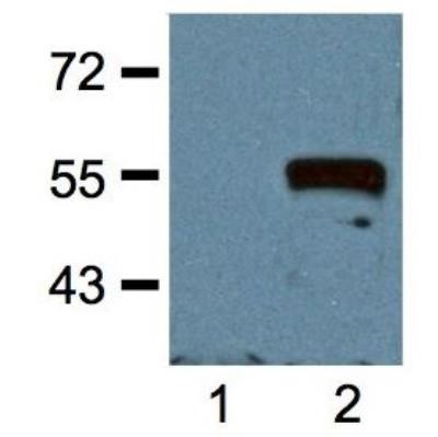
- Western Blot: c-Myc Antibody (Myc.A7) [NBP2-37822] - Analysis of 1:1000 (1ug/mL) Ab dilution probed against HEK293 cells transfected with Myc-tagged protein vector; untransfected (1) and transfected (2)
- Submitted by
- Novus Biologicals (provider)
- Main image

- Experimental details
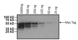
- Western Blot: c-Myc Antibody (Myc.A7) [NBP2-37822] - Analysis using the DyLight 680 conjugate of NBP2-37822. Detection of Myc Epitope Tag was performed by loading various amounts of E. coli lysate containing a multi-epitope tagged protein per well onto a 4-20% Tris-HCl polyacrylamide gel. Proteins were transferred to a PVDF membrane and blocked with 5% BSA/TBST for at least 1 hour. The membrane was probed with a DyLight 680-conjugated Myc Epitope Tag monoclonal antibody at a dilution of 1:1000 for 1 hour at room temperature on a rocking platform and washed in TBS-0.1% Tween-20. Detection was performed using the LI-COR Odyssey.

- Submitted by
- Novus Biologicals (provider)
- Main image
- Experimental details
- Western Blot: c-Myc Antibody (Myc.A7) [NBP2-37822] - Analysis using the HRP conjugate of NBP2-37822. Detection of Myc Epitope Tag was performed by loading various amounts of E. coli lysate containing a multi-epitope tagged protein per well onto a 4-20% Tris-HCl polyacrylamide gel. Proteins were transferred to a PVDF membrane and blocked with 5% BSA/TBST for at least 1 hour. The membrane was probed with an HRP-conjugated Myc Epitope Tag monoclonal antibody at a dilution of 1:1000 overnight at 4?C on a rocking platform and washed in TBS-0.1% Tween-20. Chemiluminescent detection was performed using SuperSignal West Pico.

Supportive validation
- Submitted by
- Novus Biologicals (provider)
- Main image
- Experimental details
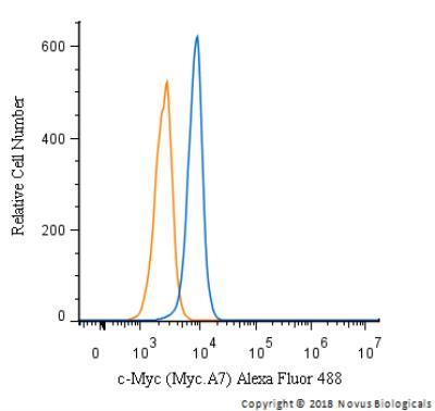
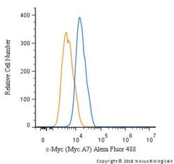
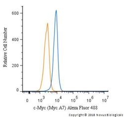
- Flow Cytometry: c-Myc Antibody (Myc.A7) [NBP2-37822] - An intracellular stain was performed on HepG2 cells with c-Myc Antibody (Myc.A7) NBP2-37822AF488 (blue) and a matched isotype control (orange). Cells were fixed with 4% PFA and then permeablized with 0.1% saponin. Cells were incubated in an antibody dilution of 10 ug/mL for 30 minutes at room temperature. Both antibodies were conjugated to Alexa Fluor 488.

- Submitted by
- Novus Biologicals (provider)
- Main image
- Experimental details
- Flow Cytometry: c-Myc Antibody (Myc.A7) [NBP2-37822] - An intracellular stain was performed on Jurkat cells with c-Myc Antibody (Myc.A7) NBP2-37822AF488 (blue) and a matched isotype control (orange). Cells were fixed with 4% PFA and then permeablized with 0.1% saponin. Cells were incubated in an antibody dilution of 10 ug/mL for 30 minutes at room temperature. Both antibodies were conjugated to Alexa Fluor 488.