 Explore
 Validate
 Learn
Learn Western blot
Antibody data
- Antibody Data
- Antigen structure
- References [2]
- Comments [0]
- Validations
- Western blot [2]
- Immunocytochemistry [3]
- Other assay [1]
Submit
Validation data
Reference
Comment
Report error
- Product number
- 710007 - Provider product page

- Provider
- Invitrogen Antibodies
- Product name
- c-Myc Recombinant Polyclonal Antibody
- Antibody type
- Polyclonal
- Antigen
- Recombinant full-length protein
- Reactivity
- Human
- Host
- Rabbit
- Isotype
- IgG
- Vial size
- 100 µg
- Concentration
- 0.5 mg/mL
- Storage
- Store at 4°C short term. For long term storage, store at -20°C, avoiding freeze/thaw cycles.
Submitted references Target guided synthesis using DNA nano-templates for selectively assembling a G-quadruplex binding c-MYC inhibitor.
Sciellin mediates mesenchymal-to-epithelial transition in colorectal cancer hepatic metastasis.
Panda D, Saha P, Das T, Dash J
Nature communications 2017 Jul 14;8:16103
Nature communications 2017 Jul 14;8:16103
Sciellin mediates mesenchymal-to-epithelial transition in colorectal cancer hepatic metastasis.
Chou CK, Fan CC, Lin PS, Liao PY, Tung JC, Hsieh CH, Hung MC, Chen CH, Chang WC
Oncotarget 2016 May 3;7(18):25742-54
Oncotarget 2016 May 3;7(18):25742-54
No comments: Submit comment
Supportive validation
- Submitted by
- Invitrogen Antibodies (provider)
- Main image

- Experimental details
- Western blot analysis of c-Myc was performed by loading 30 µg of K562, Jurkat and Raji cell lysates using Novex®NuPAGE®4-12% Bis-Tris gel (Product # NP0321BOX), Xcell SureLock Electrophoresis system (Product # EI0002), Novex sharp Pre-stained Protein Standard (Product # LC5800), and iBlot® Dry Blotting System (Product # IB21001). Proteins were transferred to a nitrocellulose membrane and blocked with 5% skim milk for 1 hour at room temperature. c-Myc was detected at ~57 kDa using c-Myc Recombinant RabbitRecombinant Rabbit Polyclonal Antibody (Product # 710007) at a 1:1000 dilution in 2.5% skim milk at 4°C overnight on a rocking platform. Detection was performed using an HRP-conjugated Goat anti-Rabbit secondary antibody (Product # G-21234) at a 1:5000 dilution and chemiluminescent detection was performed using Pierce™ ECL Western blotting Substrate (Product # 32106).

- Submitted by
- Invitrogen Antibodies (provider)
- Main image

- Experimental details
- Western blot analysis of c-Myc in Jurkat cell lysate (30 µg/lane) using a c-Myc Recombinant Rabbit Polyclonal Antibody (Product # 710007) at a dilution of 1 µg/mL. NBT/BCIP was used as the substrate (Product # WB7105).

Supportive validation
- Submitted by
- Invitrogen Antibodies (provider)
- Main image
- Experimental details
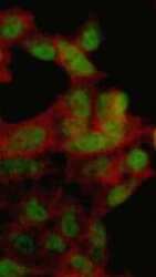
- Immunofluorescent analysis of c-Myc in HeLa cells using a c-Myc Recombinant Rabbit Polyclonal Antibody (Product # 710007) at a dilution of 10 µg/mL followed by detection using an Alexa Fluor 488-conjugated Goat anti-Rabbit secondary antibody at a dilution of 1:1000 (green) and Alexa Fluor 594 Phalloidin (red) showing predominantly nuclear cellular localization.

- Submitted by
- Invitrogen Antibodies (provider)
- Main image
- Experimental details
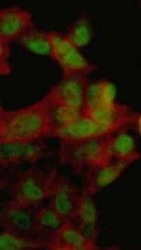
- Immunofluorescent analysis of c-Myc in HeLa cells using a c-Myc Recombinant Rabbit Polyclonal Antibody (Product # 710007) at a dilution of 10 µg/mL followed by detection using an Alexa Fluor 488-conjugated Goat anti-Rabbit secondary antibody at a dilution of 1:1000 (green) and Alexa Fluor 594 Phalloidin (red) showing predominantly nuclear cellular localization.

- Submitted by
- Invitrogen Antibodies (provider)
- Main image
- Experimental details
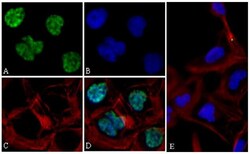
- Immunofluorescent analysis of c-Myc was performed on 70% confluent log phase U-2 OS cells. The cells were fixed with 4% paraformaldehyde for 15 minutes, permeabilized with 0. 25% Triton X-100 for 10 minutes, and blocked with 5% BSA for 1 hour at room temperature. The cells were labeled with c-Myc Recombinant Rabbit Polyclonal Antibody (Product # 710007) at a dilution of 1:400 in 1% BSA and incubated for 3 hours at room temperature and then labeled with Alexa Fluor® 488 Goat anti-Rabbit IgG secondary antibody (Product # A-11008) at a dilution of 1:400 for 30 minutes at room temperature (Panel a: green). Nuclei (Panel b: blue) were stained with SlowFade® Gold Antifade Mountant with DAPI (Product # S36938). F-actin (Panel c: red) was stained with Alexa Fluor® 594 phalloidin (Product # A12381) and panel d is a merged image showing nuclear localization. Panel e is a control without primary antibody. The images were captured using a Nikon microscope at 20X magnification.

Supportive validation
- Submitted by
- Invitrogen Antibodies (provider)
- Main image

- Experimental details
- Figure 3 SCEL activates the Wnt signaling pathway A. The protein expression of beta-catenin and its target gene c-myc was analyzed using western blot assay. beta-actin served as loading control. B. The expression of beta-catenin (green) in L1, L2, and SW620 was analyzed using confocal microscopy. DAPI (blue) was used for nuclear stain. C. and D. Western blot to determine the effect of beta-catenin or E-cadherin knockdown on EMT marker expression in L1 and L2. beta-actin served as loading control.
