Explore
 Validate
 Learn
Learn14-6784-80
antibody from Invitrogen Antibodies
Targeting: MYC
bHLHe39, c-Myc, MYCC
 Western blot Immunocytochemistry
 Immunoprecipitation Immunohistochemistry Chromatin Immunoprecipitation Other assay
Antibody data
- Antibody Data
- Antigen structure
- References [12]
- Comments [0]
- Validations
- Western blot [2]
- Immunocytochemistry [1]
- Immunohistochemistry [1]
- Chromatin Immunoprecipitation [1]
- Other assay [1]
Submit
Validation data
Reference
Comment
Report error
- Product number
- 14-6784-80 - Provider product page

- Provider
- Invitrogen Antibodies
- Product name
- c-Myc Monoclonal Antibody (9E10), eBioscience™
- Antibody type
- Monoclonal
- Antigen
- Other
- Description
- Description: The 9E10 antibody reacts with human c-Myc p67; the antibody was raised against a synthetic peptide corresponding to amino acids 408-439 within the carboxy terminal domain of human c-Myc. This antibody can be used to detect myc-tagged protein. The transcription factor c-Myc is a proto-oncogene that is at the focal point in cell cycle regulation, metabolism, apoptosis, differentiation, cell adhesion, and tumorigenesis. In normal cells the expression of c-Myc is tightly regulated but in human cancers c-Myc is frequently deregulated. C-Myc also plays a pivotal role in apoptosis, most notably its connections to the CD95/Fas death receptor pathway. These different biological responses to c-Myc are most likely the result of different overlapping subsets of c-Myc target genes. Applications Reported: This 9E10 antibody has been reported for use in immunoprecipitation, western blotting, and immunohistochemical staining of formalin-fixed paraffin embedded tissue sections. Applications Tested: This 9E10 antibody has been tested by immunohistochemistry of formalin-fixed paraffin embedded tissue using low or high pH antigen retrieval and can be used at less than or equal to 5 µg/mL. This 9E10 antibody has also been tested by western blot and can be used 5 µg/mL. It is recommended that the antibody be carefully titrated for optimal performance in the assay of interest. Purity: Greater than 90%, as determined by SDS-PAGE. Aggregation: Less than 10%, as determined by HPLC. Filtration: 0.2 µm post-manufacturing filtered.
- Reactivity
- Human
- Host
- Mouse
- Isotype
- IgG
- Antibody clone number
- 9E10
- Vial size
- 25 µg
- Concentration
- 0.5 mg/mL
- Storage
- 4° C
Submitted references Clinical-scale expansion of CD34(+) cord blood cells amplifies committed progenitors and rapid scid repopulation cells.
The NPC2 protein: A novel dog allergen.
Increased Twist expression in advanced stage of mycosis fungoides and Sézary syndrome.
Select paramyxoviral V proteins inhibit IRF3 activation by acting as alternative substrates for inhibitor of kappaB kinase epsilon (IKKe)/TBK1.
The proto-oncogene c-myc in hematopoietic development and leukemogenesis.
Translocations involving c-myc and c-myc function.
Anti-c-myc antibody 9E10: epitope key positions and variability characterized using peptide spot synthesis on cellulose.
A comprehensive system for protein purification and biochemical analysis based on antibodies to c-myc peptide.
Function of the c-Myc oncogenic transcription factor.
Mechanisms of apoptosis by c-Myc.
Epitope tag mapping of the extracellular and cytoplasmic domains of the rat parathyroid hormone (PTH)/PTH-related peptide receptor.
Ultrastructural localization of ras-related proteins using epitope-tagged plasmids.
Casamayor-Genescà A, Pla A, Oliver-Vila I, Pujals-Fonts N, Marín-Gallén S, Caminal M, Pujol-Autonell I, Carrascal J, Vives-Pi M, Garcia J, Vives J
New biotechnology 2017 Mar 25;35:19-29
New biotechnology 2017 Mar 25;35:19-29
The NPC2 protein: A novel dog allergen.
Khurana T, Newman-Lindsay S, Young PR, Slater JE
Annals of allergy, asthma & immunology : official publication of the American College of Allergy, Asthma, & Immunology 2016 May;116(5):440-446.e2
Annals of allergy, asthma & immunology : official publication of the American College of Allergy, Asthma, & Immunology 2016 May;116(5):440-446.e2
Increased Twist expression in advanced stage of mycosis fungoides and Sézary syndrome.
Goswami M, Duvic M, Dougherty A, Ni X
Journal of cutaneous pathology 2012 May;39(5):500-7
Journal of cutaneous pathology 2012 May;39(5):500-7
Select paramyxoviral V proteins inhibit IRF3 activation by acting as alternative substrates for inhibitor of kappaB kinase epsilon (IKKe)/TBK1.
Lu LL, Puri M, Horvath CM, Sen GC
The Journal of biological chemistry 2008 May 23;283(21):14269-76
The Journal of biological chemistry 2008 May 23;283(21):14269-76
The proto-oncogene c-myc in hematopoietic development and leukemogenesis.
Hoffman B, Amanullah A, Shafarenko M, Liebermann DA
Oncogene 2002 May 13;21(21):3414-21
Oncogene 2002 May 13;21(21):3414-21
Translocations involving c-myc and c-myc function.
Boxer LM, Dang CV
Oncogene 2001 Sep 10;20(40):5595-610
Oncogene 2001 Sep 10;20(40):5595-610
Anti-c-myc antibody 9E10: epitope key positions and variability characterized using peptide spot synthesis on cellulose.
Hilpert K, Hansen G, Wessner H, Küttner G, Welfle K, Seifert M, Höhne W
Protein engineering 2001 Oct;14(10):803-6
Protein engineering 2001 Oct;14(10):803-6
A comprehensive system for protein purification and biochemical analysis based on antibodies to c-myc peptide.
Hillman MC, Yang LS, Sun S, Duke JL, O'Neil KT, Kochie JE, Karjoo A, Nath P, Breth LA, Murphy K, Ross OH, Burn TC, Hollis GF, Wynn R
Protein expression and purification 2001 Nov;23(2):359-68
Protein expression and purification 2001 Nov;23(2):359-68
Function of the c-Myc oncogenic transcription factor.
Dang CV, Resar LM, Emison E, Kim S, Li Q, Prescott JE, Wonsey D, Zeller K
Experimental cell research 1999 Nov 25;253(1):63-77
Experimental cell research 1999 Nov 25;253(1):63-77
Mechanisms of apoptosis by c-Myc.
Prendergast GC
Oncogene 1999 May 13;18(19):2967-87
Oncogene 1999 May 13;18(19):2967-87
Epitope tag mapping of the extracellular and cytoplasmic domains of the rat parathyroid hormone (PTH)/PTH-related peptide receptor.
Xie LY, Abou-Samra AB
Endocrinology 1998 Nov;139(11):4563-7
Endocrinology 1998 Nov;139(11):4563-7
Ultrastructural localization of ras-related proteins using epitope-tagged plasmids.
Robertson D, Paterson HF, Adamson P, Hall A, Monaghan P
The journal of histochemistry and cytochemistry : official journal of the Histochemistry Society 1995 May;43(5):471-80
The journal of histochemistry and cytochemistry : official journal of the Histochemistry Society 1995 May;43(5):471-80
No comments: Submit comment
Supportive validation
- Submitted by
- Invitrogen Antibodies (provider)
- Main image
- Experimental details
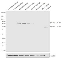
- Western blot was performed using Anti-Myc Tag Monoclonal Antibody (Product # 14-6784-82) by loading whole cell extracts of untransfected and transiently transfected HEK-293E lysates: untransfected, 60 µg (Lane 1), empty vector control, 60 µg (Lane 2), Myc-p65-V5, 60 µg (Lane 3), Myc-p65-V5, 40 µg (Lane 4), Myc-p65-V5, 20 µg (Lane 5), Myc-p65-V5, 10 µg (Lane 6), p65-HA, 60 µg (Lane 7) and 25 ng of Positope (Lane 9) were electrophoresed using NuPAGE™ 4-12% Bis-Tris Protein Gel (Product # NP0321BOX). Resolved proteins were then transferred onto a nitrocellulose membrane (Product # IB23001) by iBlot® 2 Dry Blotting System (Product # IB21001). A ~65 kDa band corresponding to Myc-p65-V5 was observed in HEK293E transfected lysates on probing with the primary antibody (1 µg/mL) and detected by chemiluminescence with Goat anti-Mouse IgG (H+L) Superclonal™ Secondary Antibody, HRP (Product # A28177, 1:4000 dilution). Positope (Product # R90050). Positope (Product # R90050) is a 53 kDa recombinant protein consisting multiple epitope tags, which has been used as a positive control for Myc detection. No cross-reactivity was seen with p65-HA expressing lysate.

- Submitted by
- Invitrogen Antibodies (provider)
- Main image
- Experimental details
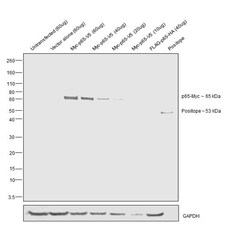
- Western Blot was performed using Anti-Myc Tag Monoclonal Antibody (Product # 14-6784-82) by loading whole cell extracts of untransfected and transiently transfected HEK-293E lysates: untransfected, 60 µg (Lane 1), empty vector control, 60 µg (Lane 2), Myc-p65-V5, 60 µg (Lane 3), Myc-p65-V5, 40 µg (Lane 4), Myc-p65-V5, 20 µg (Lane 5), Myc-p65-V5, 10 µg (Lane 6), FLAG-p65-HA, 60 µg (Lane 7) and 25 ng of Positope (Lane 8) were electrophoresed using NuPAGE™ 4-12% Bis-Tris Protein Gel (Product # NP0321BOX). Resolved proteins were then transferred onto a nitrocellulose membrane (Product # IB23001) by iBlot® 2 Dry Blotting System (Product # IB21001). A ~65 kDa band corresponding to Myc-p65-V5 was observed in HEK293E transfected lysates on probing with the primary antibody (1 µg/mL) and detected by chemiluminescence with Goat anti-Mouse IgG (H+L) Superclonal™ Secondary Antibody, HRP (Product # A28177) . Chemiluminescent detection was performed using Novex® ECL Chemiluminescent Substrate Reagent Kit (Product # WP20005). Positope (Product # R90050). Positope (Product # R90050) is a 53 kDa recombinant protein consisting multiple epitope tags, which has been used as a positive control for Myc detection. No cross-reactivity was seen with FLAG-HA-tagged p65 expressing lysate.

Supportive validation
- Submitted by
- Invitrogen Antibodies (provider)
- Main image
- Experimental details
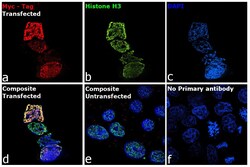
- Immunofluorescent analysis of Myc tag was performed using 70% confluent His-H3-Myc transfected HEK-293 cells. The cells were fixed with 4% paraformaldehyde for 10 minutes, permeabilized with 0.1% Triton™ X-100 for 15 minutes, and blocked with 2% BSA for 1 hour at room temperature. The cells were labeled with c-Myc Monoclonal Antibody (9E10) (Product # 14-6784-82) at 1:100 dilution and Histone H3 antibody (Product # 711055) at 1:200 dilution in 0.1% BSA, incubated at 4 degree Celsius overnight and then labeled with Goat anti-Rabbit IgG (H+L) Highly Cross-Adsorbed Secondary Antibody, Alexa Fluor Plus 647 (Product # A32733) respectively at a dilution of 1:2000 for 45 minutes at room temperature. Panel a (Nuclei: Red) represents Myc tag. Panel b (Nuclei: Green) represents Histone 3. Panel c (Nuclei: Blue) represents ProLong™ Diamond Antifade Mountant with DAPI (Product # P36962). Panel d represents the merged image showing the co-localization of nuclear signals in transfected cells. Panel e represents un-transfected HEK-293 cells. Panel f represents isotype control cells to assess background. The images were captured at 60X magnification.

Supportive validation
- Submitted by
- Invitrogen Antibodies (provider)
- Main image
- Experimental details
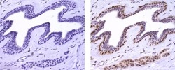
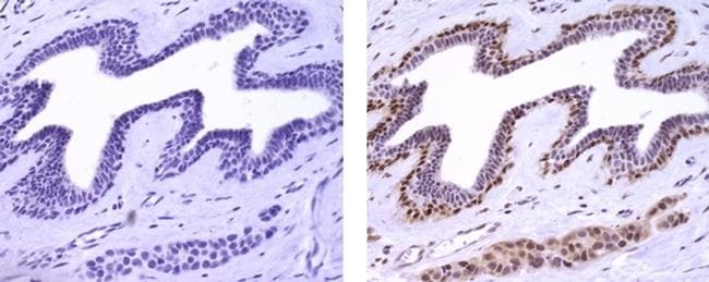
- Immunohistochemistry of formalin-fixed paraffin embedded human breast carcinoma using 5 µg/mL Mouse IgG1 K Isotype Control Purified (left) or 5 µg/mL Anti-Human c-Myc p67 Purified (right), followed by Anti-Mouse IgG Biotin, Streptavidin HRP, and DAB visualization.Nuclei are counterstained with hematoxylin.
Supportive validation
- Submitted by
- Invitrogen Antibodies (provider)
- Main image

- Experimental details
- HEK293E cells were either untransfected or transfected with an Myc-Tagged Human H3 construct. Chromatin Immunoprecipitation (ChIP) was performed using target H3 antibody (Product # 711055) or c-Myc Monoclonal Antibody (9E10) (Product # 14-6784-82, 5 µg) on sheared chromatin from untransfected and myc tag transfected HEK293E cells using the MAGnify ChIP System kit (Product # 49-2024). Normal Rabbit IgG was used as a negative IP control for target H3 antibody and Normal Mouse IgG for Myc Tag antibody. The purified DNA was analyzed by qPCR using primers binding to RPL30, ALDOA, PABPC1, cFOS and MYOD. Data is presented as fold enrichment of the antibody signal in untransfected and Myc tagged Human H3 construct transfected cells versus the negative control IgG using the comparative CT method.

Supportive validation
- Submitted by
- Invitrogen Antibodies (provider)
- Main image

- Experimental details
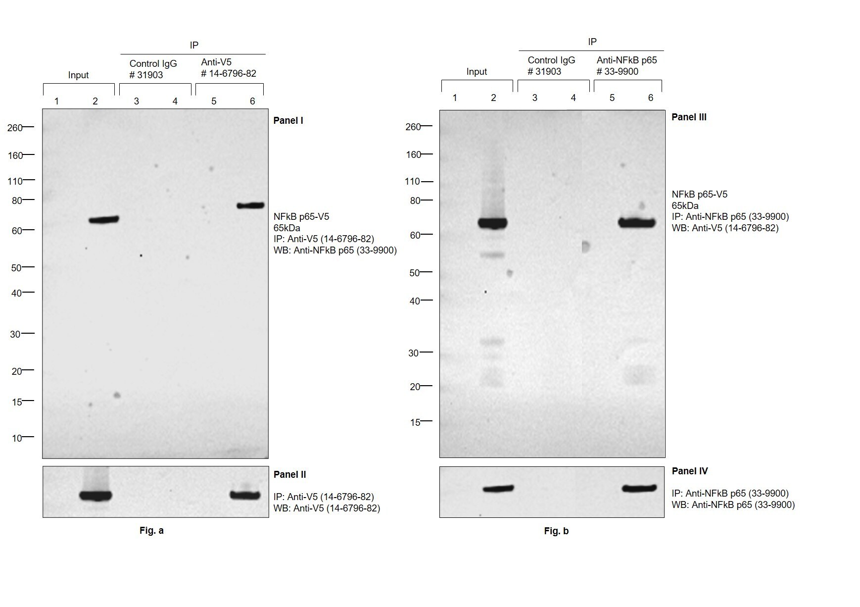
- Immunoprecipitation (IP) studies of V5 tag were performed with V5 Tag Monoclonal Antibody (TCM5), eBioscience™ (Product # 14-6796-82) (Panel I, II) or NFkB p65 Monoclonal Antibody (Product # 33-9900) (Panel III, IV) using the Dynabeads® Protein G Immunoprecipitation Kit (Product # 10007D). Lane 1,2: Total cell extract of HEK-293E untransfected (UT) and transfected (T) with V5-tagged NFkB p65 construct (7% of input) Lane 3,4: IP of UT and T HEK-293E lysate using Isotype-matched Mouse IgG (Product # 31903) Lane 5,6: IP of UT and T HEK-293E lysate with 5ug of V5 Tag Monoclonal Antibody (TCM5), eBioscience™ (Product # 14-6796-82) (Panel I, II) or NFkB p65 Monoclonal Antibody (Product # 33-9900) (Panel III, IV) IP was analyzed using western blot. V5-tagged NFkB p65 was detected at ~65 kDa by probing the blot with NFkB p65 Monoclonal Antibody (Product # 33-9900) (0.5 µg/mL dilution) (Panel I, IV) or V5 Tag Monoclonal Antibody (TCM5), eBioscience™ (Product # 14-6796-82) (1:1,000 dilution) (Panel II, III) and detected by chemiluminescence with Recombinant Protein G-HRP (Product # 101223) (1:500 dilution) using the iBright FL 1500 (Product # A44241). Chemiluminescent detection was performed using Novex® ECL Chemiluminescent Substrate Reagent Kit (Product # WP20005).