 Explore
 Validate
 Learn
Learn Western blot
 Immunocytochemistry
Antibody data
- Antibody Data
- Antigen structure
- References [6]
- Comments [0]
- Validations
- Immunocytochemistry [2]
- Immunoprecipitation [1]
- Chromatin Immunoprecipitation [1]
- Other assay [1]
Submit
Validation data
Reference
Comment
Report error
- Product number
- PA1-981 - Provider product page

- Provider
- Invitrogen Antibodies
- Product name
- Anti-Myc Tag Polyclonal Antibody
- Antibody type
- Polyclonal
- Antigen
- Synthetic peptide
- Description
- PA1-981 detects c-Myc tagged proteins. PA1-981 has been successfully used in WB and immunofluorescence procedures to detect the presence of expressed c-Myc tagged proteins in transfected mammalian cells. PA1-981 immunogen is a synthetic peptide corresponding to residues C E(410) Q K L I S E E D L(419) of human c-Myc. PA1-981 immunizing peptide (PEP-035) is available for use in neutralization and control experiments.
- Host
- Rabbit
- Isotype
- IgG
- Vial size
- 100 µL
- Concentration
- 1 mg/mL
- Storage
- -20° C, Avoid Freeze/Thaw Cycles
Submitted references Suppression and synthetic-lethal genetic relationships of ΔgpsB mutations indicate that GpsB mediates protein phosphorylation and penicillin-binding protein interactions in Streptococcus pneumoniae D39.
Mechanism of early light signaling by the carboxy-terminal output module of Arabidopsis phytochrome B.
Identification of Interferon-Stimulated Gene Proteins That Inhibit Human Parainfluenza Virus Type 3.
Unique nonstructural proteins of Pneumonia Virus of Mice (PVM) promote degradation of interferon (IFN) pathway components and IFN-stimulated gene proteins.
Excessive sarcoplasmic/endoplasmic reticulum Ca2+-ATPase expression causes increased sarcoplasmic reticulum Ca2+ uptake but decreases myocyte shortening.
Increased expression of the glial glutamate transporter EAAT2 modulates excitotoxicity and delays the onset but not the outcome of ALS in mice.
Rued BE, Zheng JJ, Mura A, Tsui HT, Boersma MJ, Mazny JL, Corona F, Perez AJ, Fadda D, Doubravová L, Buriánková K, Branny P, Massidda O, Winkler ME
Molecular microbiology 2017 Mar;103(6):931-957
Molecular microbiology 2017 Mar;103(6):931-957
Mechanism of early light signaling by the carboxy-terminal output module of Arabidopsis phytochrome B.
Qiu Y, Pasoreck EK, Reddy AK, Nagatani A, Ma W, Chory J, Chen M
Nature communications 2017 Dec 4;8(1):1905
Nature communications 2017 Dec 4;8(1):1905
Identification of Interferon-Stimulated Gene Proteins That Inhibit Human Parainfluenza Virus Type 3.
Rabbani MA, Ribaudo M, Guo JT, Barik S
Journal of virology 2016 Dec 15;90(24):11145-11156
Journal of virology 2016 Dec 15;90(24):11145-11156
Unique nonstructural proteins of Pneumonia Virus of Mice (PVM) promote degradation of interferon (IFN) pathway components and IFN-stimulated gene proteins.
Dhar J, Barik S
Scientific reports 2016 Dec 1;6:38139
Scientific reports 2016 Dec 1;6:38139
Excessive sarcoplasmic/endoplasmic reticulum Ca2+-ATPase expression causes increased sarcoplasmic reticulum Ca2+ uptake but decreases myocyte shortening.
Teucher N, Prestle J, Seidler T, Currie S, Elliott EB, Reynolds DF, Schott P, Wagner S, Kogler H, Inesi G, Bers DM, Hasenfuss G, Smith GL
Circulation 2004 Dec 7;110(23):3553-9
Circulation 2004 Dec 7;110(23):3553-9
Increased expression of the glial glutamate transporter EAAT2 modulates excitotoxicity and delays the onset but not the outcome of ALS in mice.
Guo H, Lai L, Butchbach ME, Stockinger MP, Shan X, Bishop GA, Lin CL
Human molecular genetics 2003 Oct 1;12(19):2519-32
Human molecular genetics 2003 Oct 1;12(19):2519-32
No comments: Submit comment
Supportive validation
- Submitted by
- Invitrogen Antibodies (provider)
- Main image
- Experimental details
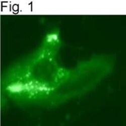
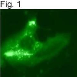
- Immunolocalization of c-Myc in transfected mammalian cells using Product # PA1-981.

- Submitted by
- Invitrogen Antibodies (provider)
- Main image
- Experimental details
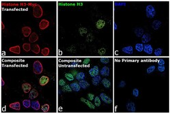
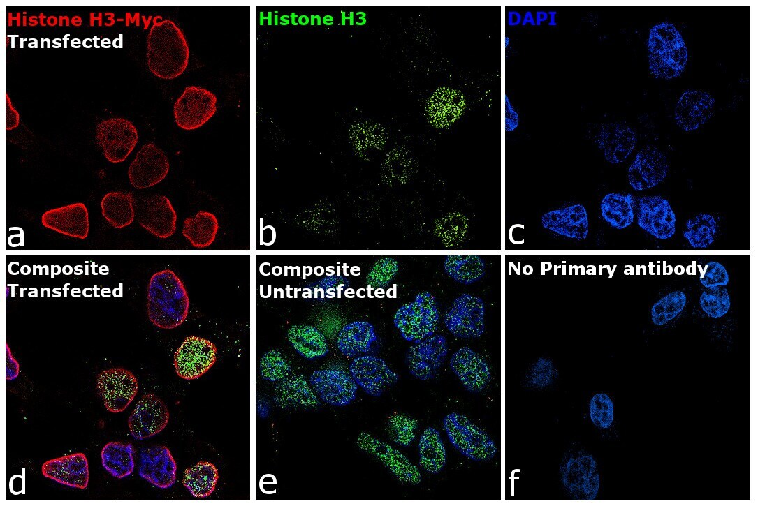
- Immunofluorescence analysis of Myc proto-oncogene protein was performed using 70% confluent log phase HEK-293 cells. The cells were fixed with 4% paraformaldehyde for 10 minutes, permeabilized with 0.1% Triton™ X-100 for 15 minutes, and blocked with 2% BSA for 45 minutes at room temperature. The cells were labeled with Myc Tag Polyclonal Antibody (Product # PA1-981) at 1:100 in 0.1% BSA and Histone H3 antibody (Product # AHO1432) at 1:200 dilution in 0.1% BSA, incubated at 4 degree Celsius overnight and then labeled with Goat anti-Rabbit IgG (H+L) Highly Cross-Adsorbed Secondary Antibody, Alexa Fluor Plus 647 (Product # A32733) and Goat anti-Mouse IgG Alexa Fluor Plus 488 (Product # A21042) respectively at a dilution of 1:2,000 for 45 minutes at room temperature. Panel a (Nuclei: Red) represents Myc tag. Panel b (Nuclei: Green) represents Histone 3. Panel c (Nuclei: Blue) represents ProLong™ Diamond Antifade Mountant with DAPI (Product # P36962). Panel d represents the merged image showing the co-localization of nuclear signals in transfected cells. Panel e represents un-transfected HEK-293 cells. Panel f represents control cells with no primary antibody to assess background. The images were captured at 60X magnification.
Supportive validation
- Submitted by
- Invitrogen Antibodies (provider)
- Main image
- Experimental details
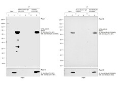
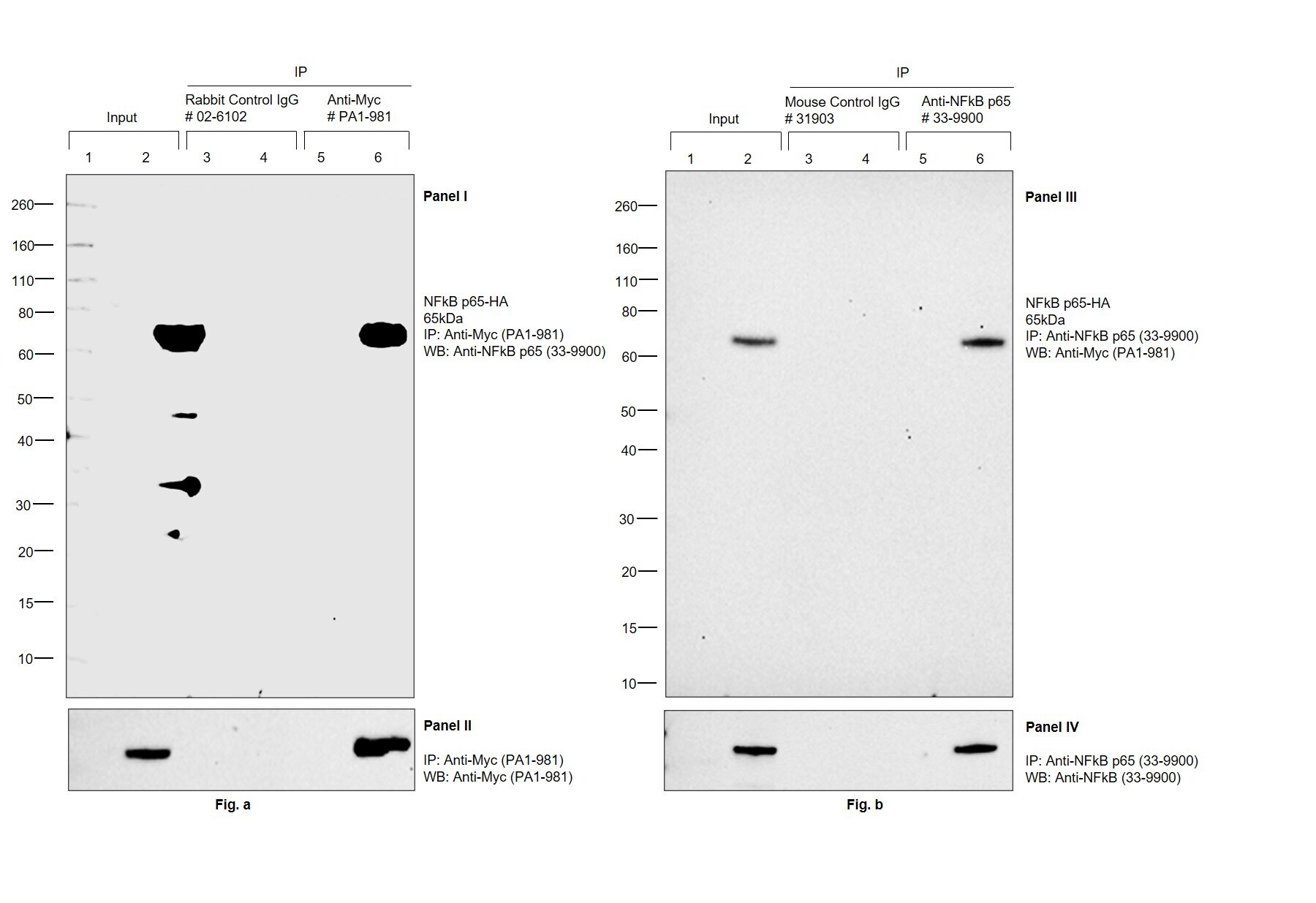
- Immunoprecipitation (IP) studies of Myc tag were performed with Myc Tag Polyclonal Antibody (Product # PA1-981) (Panel I, II) or NFkB p65 Monoclonal Antibody (Product # 33-9900) (Panel III, IV) using the Dynabeads® Protein A Immunoprecipitation Kit (Product # 10006D) and Dynabeads® Protein G Immunoprecipitation Kit (Product # 10007D) respectively. Lane 1,2: Total cell extract of HEK-293E untransfected (UT) and transfected (T) with Myc-tagged NFkB p65 construct (7% of input) Lane 3,4: IP of UT and T HEK-293E lysate using Isotype-matched Rabbit IgG (Product # 02-6102) (Panel I,II) and Isotype-matched Mouse IgG (Product # 31903) (Panel III,IV) Lane 5,6: IP of UT and T HEK-293E lysate with 5 µg of Myc Tag Polyclonal Antibody (Product # PA1-981) (Panel I, II) or NFkB p65 Monoclonal Antibody (Product # 33-9900) (Panel III, IV) IP was analyzed using western blot. HA-tagged NFkB p65 was detected at ~65 kDa by probing the blot with NFkB p65 Monoclonal Antibody (Product # 33-9900) (0.5 µg/mL dilution) (Panel I, IV) or Myc Tag Polyclonal Antibody (Product # PA1-981) (1:1,000 dilution) (Panel II, III) and detected by chemiluminescence with Clean-Blot™ IP Detection Kit (HRP) (Product # 21232) (Panel II,III) and Recombinant Protein G-HRP (Product # 101223) (1:500 dilution) (Panel I, IV) using the iBright FL 1500 (Product # A44241). Chemiluminescent detection was performed using Novex® ECL Chemiluminescent Substrate Reagent Kit (# WP20005).
Supportive validation
- Submitted by
- Invitrogen Antibodies (provider)
- Main image
- Experimental details
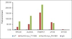
- HEK293E cells were either untransfected or transfected with an Myc-Tagged Human H3 construct. Chromatin Immunoprecipitation (ChIP) was performed using target H3 antibody (Product # 711055) or Myc Tag Polyclonal Antibody (Product # PA1-981, 5 µg) on sheared chromatin from untransfected and Myc tag transfected HEK293E cells using the MAGnify ChIP System kit (Product # 49-2024). Normal Rabbit IgG was used as a negative IP control. The purified DNA was analyzed by qPCR using primers binding to RPL30, ALDOA, PABPC1, cFOS and MYOD. Data is presented as fold enrichment of the antibody signal in untransfected and Myc Tagged Human H3 construct transfected cells versus the negative control IgG using the comparative CT method.

Supportive validation
- Submitted by
- Invitrogen Antibodies (provider)
- Main image
- Experimental details
- Immunolocalization of c-Myc in transfected mammalian cells using Product # PA1-981.
