Explore
 Validate
 Learn
Learn Western blot
 Immunohistochemistry
Antibody data
- Antibody Data
- Antigen structure
- References [2]
- Comments [0]
- Validations
- Western blot [4]
- Immunocytochemistry [2]
- Chromatin Immunoprecipitation [1]
Submit
Validation data
Reference
Comment
Report error
- Product number
- AF3696 - Provider product page

- Provider
- R&D Systems
- Product name
- Human/Mouse c-Myc Antibody
- Antibody type
- Polyclonal
- Description
- Antigen Affinity-purified. Detects human c-Myc in direct ELISAs. Detects human and mouse c-Myc in Western blots. In direct ELISAs, less than 1% cross-reactivity with recombinant human (rh) L-Myc and rhN-Myc is observed.
- Reactivity
- Human, Mouse
- Host
- Goat
- Conjugate
- Unconjugated
- Antigen sequence
P01106- Isotype
- IgG
- Vial size
- 100 ug
- Concentration
- LYOPH
- Storage
- Use a manual defrost freezer and avoid repeated freeze-thaw cycles. 12 months from date of receipt, -20 to -70 °C as supplied. 1 month, 2 to 8 °C under sterile conditions after reconstitution. 6 months, -20 to -70 °C under sterile conditions after reconstitution.
Submitted references Lysine-Specific Demethylase 1 (LSD1/KDM1A) Is a Novel Target Gene of c-Myc.
Convergence of cMyc and β-catenin on Tcf7l1 enables endoderm specification.
Nagasaka M, Tsuzuki K, Ozeki Y, Tokugawa M, Ohoka N, Inoue Y, Hayashi H
Biological & pharmaceutical bulletin 2019;42(3):481-488
Biological & pharmaceutical bulletin 2019;42(3):481-488
Convergence of cMyc and β-catenin on Tcf7l1 enables endoderm specification.
Morrison G, Scognamiglio R, Trumpp A, Smith A
The EMBO journal 2016 Feb 1;35(3):356-68
The EMBO journal 2016 Feb 1;35(3):356-68
No comments: Submit comment
Supportive validation
- Submitted by
- R&D Systems (provider)
- Main image
- Experimental details
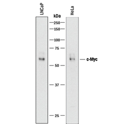
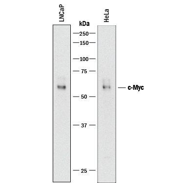
- Detection of Human c-Myc by Western Blot. Western blot shows lysates of LNCaP human prostate cancer cell line and HeLa human cervical epithelial carcinoma cell line. PVDF membrane was probed with 0.5 µg/mL of Goat Anti-Human/Mouse c-Myc Antigen Affinity-purified Polyclonal Antibody (Catalog # AF3696) followed by HRP-conjugated Anti-Goat IgG Secondary Antibody (Catalog # HAF017). A specific band was detected for c-Myc at approximately 56 kDa (as indicated). This experiment was conducted under reducing conditions and using Immunoblot Buffer Group 1.
- Submitted by
- R&D Systems (provider)
- Main image

- Experimental details
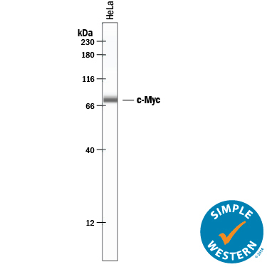
- Detection of Human c-Myc by Simple WesternTM. Simple Western lane view shows lysates of HeLa human cervical epithelial carcinoma cell line, loaded at 0.2 mg/mL. A specific band was detected for c-Myc at approximately 78 kDa (as indicated) using 20 µg/mL of Goat Anti-Human/Mouse c-Myc Antigen Affinity-purified Polyclonal Antibody (Catalog # AF3696) followed by 1:50 dilution of HRP-conjugated Anti-Goat IgG Secondary Antibody (Catalog # HAF109). This experiment was conducted under reducing conditions and using the 12-230 kDa separation system.
- Submitted by
- R&D Systems (provider)
- Main image

- Experimental details
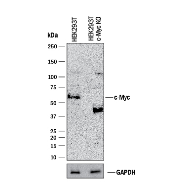
- Western Blot Shows Human c-Myc Specificity by Using Knockout Cell Line. Western blot shows lysates of HEK293T human embryonic kidney parental cell line and c-Myc knockout HEK293T cell line (KO). PVDF membrane was probed with 0.5 µg/mL of Goat Anti-Human/Mouse c-Myc Antigen Affinity-purified Polyclonal Antibody (Catalog # AF3696) followed by HRP-conjugated Anti-Goat IgG Secondary Antibody (Catalog # HAF017). A specific band was detected for c-Myc at approximately 52 kDa (as indicated) in the parental HEK293T cell line, but is not detectable in knockout HEK293T cell line. GAPDH (Catalog # AF5718) is shown as a loading control. This experiment was conducted under reducing conditions and using Immunoblot Buffer Group 1.
- Submitted by
- R&D Systems (provider)
- Main image
- Experimental details
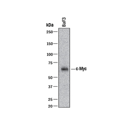
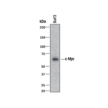
- Detection of Mouse c-Myc by Western Blot. Western blot shows lysates of BaF3 mouse pro-B cell line. PVDF membrane was probed with 0.5 µg/mL of Goat Anti-Human/Mouse c-Myc Antigen Affinity-purified Polyclonal Antibody (Catalog # AF3696) followed by HRP-conjugated Anti-Goat IgG Secondary Antibody (Catalog # HAF017). A specific band was detected for c-Myc at approximately 56 kDa (as indicated). This experiment was conducted under reducing conditions and using Immunoblot Buffer Group 1.
Supportive validation
- Submitted by
- R&D Systems (provider)
- Main image

- Experimental details
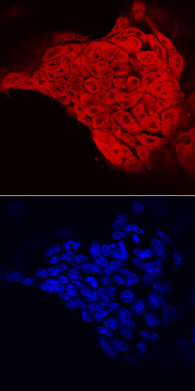
- c-Myc in D3 Mouse Stem Cells. c-Myc was detected in immersion fixed D3 mouse embryonic stem cell line using Goat Anti-Human/Mouse c-Myc Antigen Affinity-purified Polyclonal Antibody (Catalog # AF3696) at 10 µg/mL for 3 hours at room temperature. Cells were stained using the NorthernLights™ 557-conjugated Anti-Goat IgG Secondary Antibody (red, upper panel; Catalog # NL001) and counterstained with DAPI (blue, lower panel). Specific staining was localized to nuclei and cytoplasm. View our protocol for Fluorescent ICC Staining of Cells on Coverslips.
- Submitted by
- R&D Systems (provider)
- Main image

- Experimental details
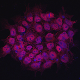
- c-Myc in BG01V Human Stem Cells. c-Myc was detected in immersion fixed BG01V human embryonic stem cells using 10 µg/mL Goat Anti-Human/Mouse c-Myc Antigen Affinity-purified Polyclonal Antibody (Catalog # AF3696) for 3 hours at room temperature. Cells were stained with the NorthernLights™ 557-conjugated Anti-Goat IgG Secondary Antibody (red; Catalog # NL001) and counterstained with DAPI (blue). View our protocol for Fluorescent ICC Staining of Cells on Coverslips.
Supportive validation
- Submitted by
- R&D Systems (provider)
- Main image

- Experimental details
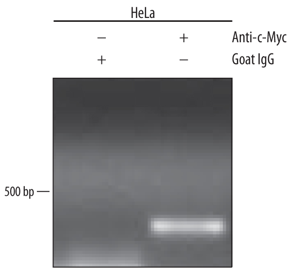
- Detection of c-Myc-regulated Genes by Chromatin Immunoprecipitation. HeLa human cervical epithelial carcinoma cell line was fixed using formaldehyde, resuspended in lysis buffer, and sonicated to shear chromatin. c-Myc/DNA complexes were immunoprecipitated using 5 μg Goat Anti-Human/Mouse c-Myc Antigen Affinity-purified Polyclonal Antibody (Catalog # AF3696) or control antibody (Catalog # AB-108-C) for 15 minutes in an ultrasonic bath, followed by Biotinylated Anti-Goat IgG Secondary Antibody (Catalog # BAF109). Immunocomplexes were captured using 50 μL of MagCellect Streptavidin Ferrofluid (Catalog # MAG999) and DNA was purified using chelating resin solution. The p21 promoter was detected by standard PCR.