Explore
 Validate
 Learn
Learn Western blot
 Immunocytochemistry
Antibody data
- Antibody Data
- Antigen structure
- References [1]
- Comments [0]
- Validations
- Western blot [1]
- Immunocytochemistry [1]
- Immunohistochemistry [1]
Submit
Validation data
Reference
Comment
Report error
- Product number
- AMAb90956 - Provider product page

- Provider
- Atlas Antibodies
- Proper citation
- Atlas Antibodies Cat#AMAb90956, RRID:AB_2665731
- Product name
- Anti-p53
- Antibody type
- Monoclonal
- Description
- Monoclonal Antibody against Human TP53, Clone ID: CL2199, Gene description: tumor protein p53, Alternative Gene Names: TP53, LFS1, Validated applications: WB, IHC, ICC, Uniprot ID: P04637, Storage: Store at +4°C for short term storage. Long time storage is recommended at -20°C.
- Reactivity
- Human
- Host
- Mouse
- Conjugate
- Unconjugated
- Isotype
- IgG
- Antibody clone number
- CL2199
- Vial size
- 100 µl
- Concentration
- 1.0 mg/ml
- Storage
- Store at +4°C for short term storage. Long time storage is recommended at -20°C.
- Handling
- The antibody solution should be gently mixed before use.
Submitted references Downregulation of the cancer susceptibility protein WRAP53β in epithelial ovarian cancer leads to defective DNA repair and poor clinical outcome.
Hedström E, Pederiva C, Farnebo J, Nodin B, Jirström K, Brennan DJ, Farnebo M
Cell death & disease 2015 Oct 1;6(10):e1892
Cell death & disease 2015 Oct 1;6(10):e1892
No comments: Submit comment
Enhanced validation
- Submitted by
- Atlas Antibodies (provider)
- Enhanced method
- Genetic validation
- Main image

- Experimental details
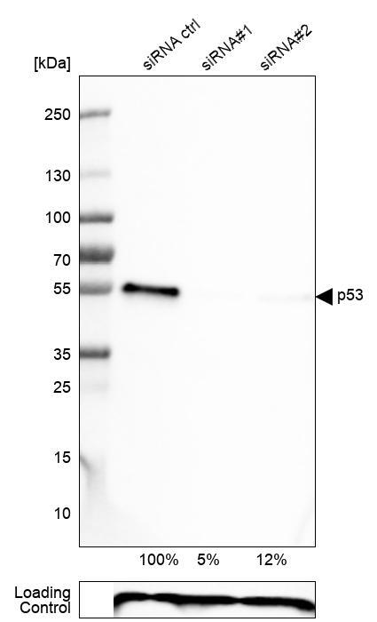
- Western blot analysis in U-251MG cells transfected with control siRNA, target specific siRNA probe #1 and #2, using Anti-p53 antibody. Remaining relative intensity is presented. Loading control: Anti-PPIB.
- Sample type
- Human
- Protocol
- Protocol
Supportive validation
- Submitted by
- Atlas Antibodies (provider)
- Main image
- Experimental details
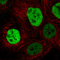
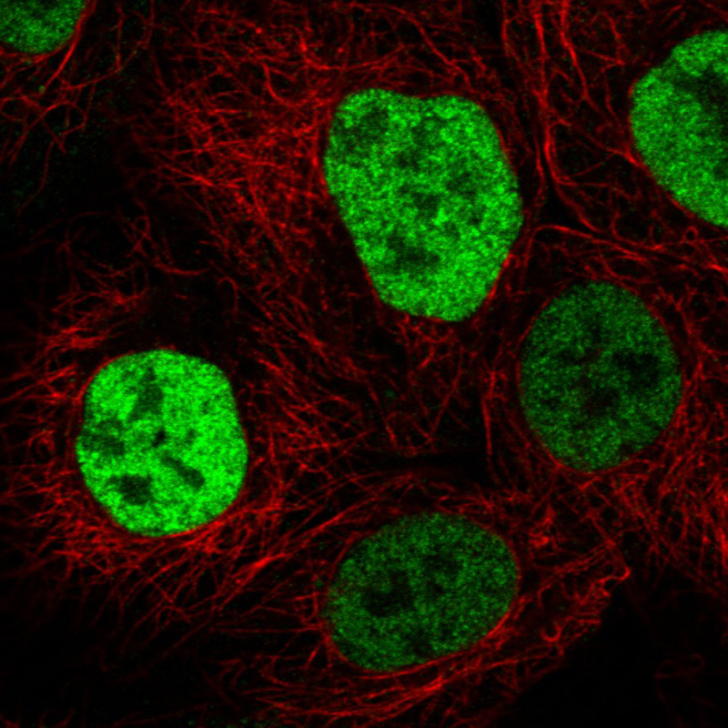
- Immunofluorescence staining in A431 cell line with Anti-p53 monoclonal antibody, showing cell cycle dependent nuclear (without nucleoli) staining in green. Microtubule- and nuclear probes are visualized in red and blue respectively (where available).
- Sample type
- Human
Supportive validation
- Submitted by
- Atlas Antibodies (provider)
- Enhanced method
- Orthogonal validation
- Main image
- Experimental details
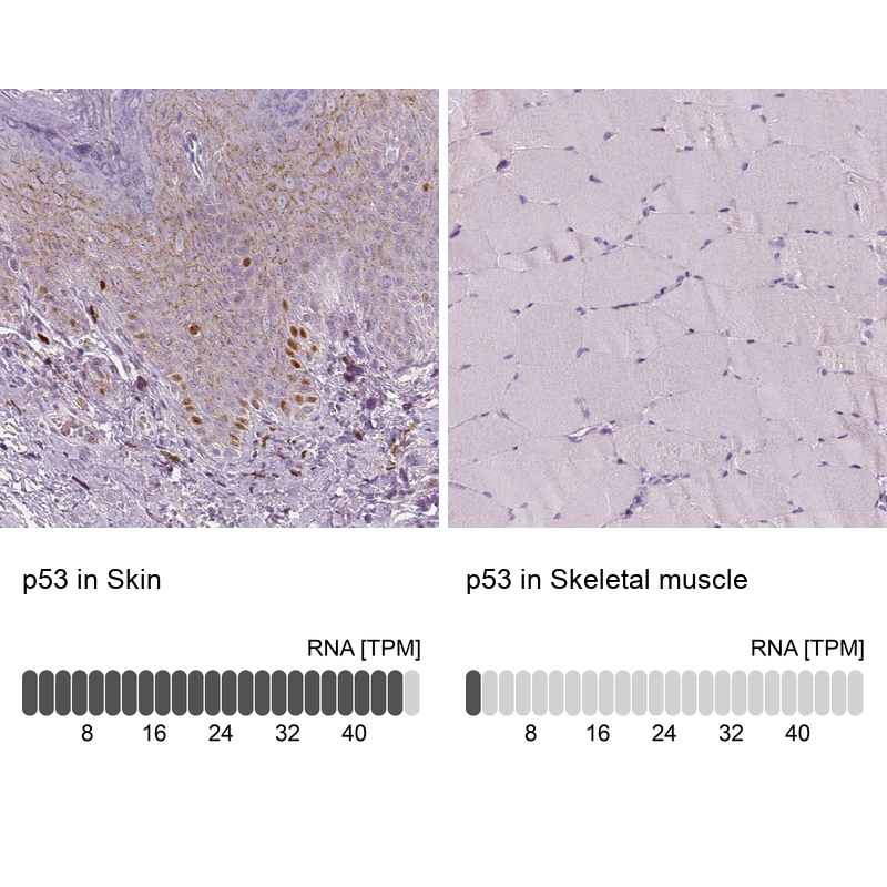
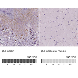
- Immunohistochemistry analysis in human skin and skeletal muscle tissues using AMAb90956 antibody. Corresponding p53 RNA-seq data are presented for the same tissues.
- Sample type
- Human
- Protocol
- Protocol