 Explore
 Validate
 Learn
Learn Western blot
Antibody data
- Antibody Data
- Antigen structure
- References [0]
- Comments [0]
- Validations
- Western blot [3]
- Immunocytochemistry [1]
- Immunohistochemistry [3]
- Chromatin Immunoprecipitation [1]
Submit
Validation data
Reference
Comment
Report error
- Product number
- 710294 - Provider product page

- Provider
- Invitrogen Antibodies
- Product name
- Acetyl-p53 (Lys382) Recombinant Polyclonal Antibody (10HCLC)
- Antibody type
- Polyclonal
- Antigen
- Synthetic peptide
- Reactivity
- Human, Mouse
- Host
- Rabbit
- Isotype
- IgG
- Antibody clone number
- 10HCLC
- Vial size
- 100 µg
- Concentration
- 0.5 mg/mL
- Storage
- Store at 4°C short term. For long term storage, store at -20°C, avoiding freeze/thaw cycles.
No comments: Submit comment
Supportive validation
- Submitted by
- Invitrogen Antibodies (provider)
- Main image
- Experimental details
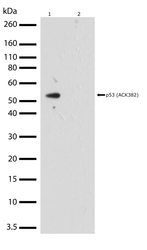
- Western blot analysis of p53 in whole cell extracts from HeLa cells treated with doxorubicin (0.2 uM) and sodium butyrate (5mM, 24 hrs) (lane 1) or lysates from untreated HeLa cells (lane 2) using a p53 Recombinant Rabbit Polyclonal Antibody (Product # 710294) at a dilution of 2 µg/mL. Samples were detected using chemiluminescence (ECL). Results show a band at ~53kDa.

- Submitted by
- Invitrogen Antibodies (provider)
- Main image

- Experimental details
- Western blot analysis of p53 in whole cell extracts from HeLa cells treated with doxorubicin (0.2 uM) and sodium butyrate (5mM, 24 hrs) (lane 1) or lysates from untreated HeLa cells (lane 2) using a p53 Recombinant Rabbit Polyclonal Antibody (Product # 710294) at a dilution of 2 µg/mL. Samples were detected using chemiluminescence (ECL). Results show a band at ~53kDa.

- Submitted by
- Invitrogen Antibodies (provider)
- Main image
- Experimental details
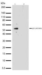
- Western blot analysis was performed on whole cell extracts from Hela treated with Doxorubicin (treated at 0.2 µM for 24hrs) and Sodium butyrate (treated at 5mM for 24hrs) (Lane 1) and Hela lysate without treatemnt (Lane 2, negative control). Endogenous level of acetylated P53 (AcK382) was detected at ~53 kDa was detected using P53 (AcK382) Recombinant Rabbit Polyclonal Antibody (Product # 710294) at 2 µg/mL. The blot was developed using chemiluminescence (ECL) method.

Supportive validation
- Submitted by
- Invitrogen Antibodies (provider)
- Main image

- Experimental details
- Immunofluorescent analysis of Acetyl p53 Lys382 in HeLa cells using an Acetyl p53 Lys382 Recombinant Rabbit Polyclonal Antibody (Product # 710294) followed by detection using an Alexa Fluor 488-conjugated Goat anti-Rabbit secondary antibody (green) (Image A). Nuclei were stained using DAPI (Image B) and actin stained with Alexa Fluor 594 phalloidin (red) (image C). Image D is a composite image showing subcellular cytoplasmic localization of acetylated p53 and image E is a composite image showing specificity of antibody to acetylated p53 by lack of staining in untreated cells.

Supportive validation
- Submitted by
- Invitrogen Antibodies (provider)
- Main image
- Experimental details
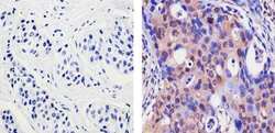
- Immunohistochemistry analysis of P53 (ACK 382) showing staining in the cytoplasm and nucleus of paraffin-embedded human breast carcinoma (right) compared to a negative control without primary antibody (left). To expose target proteins, antigen retrieval was performed using 10mM sodium citrate (pH 6.0), microwaved for 8-15 min. Following antigen retrieval, tissues were blocked in 3% H2O2-methanol for 15 min at room temperature, washed with ddH2O and PBS, and then probed with a P53 (ACK 382) Recombinant Rabbit Polyclonal Antibody (Product # 710294) diluted in 3% BSA-PBS at a dilution of 1:20 for 1 hour at 37ºC in a humidified chamber. Tissues were washed extensively in PBST and detection was performed using an HRP-conjugated secondary antibody followed by colorimetric detection using a DAB kit. Tissues were counterstained with hematoxylin and dehydrated with ethanol and xylene to prep for mounting.

- Submitted by
- Invitrogen Antibodies (provider)
- Main image
- Experimental details
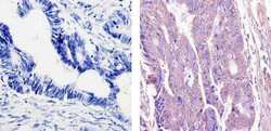
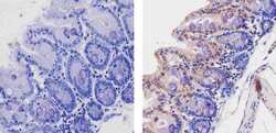
- Immunohistochemistry analysis of P53 (ACK 382) showing staining in the cytoplasm and weak staining in the nucleus of paraffin-embedded human colon carcinoma (right) compared to a negative control without primary antibody (left). To expose target proteins, antigen retrieval was performed using 10mM sodium citrate (pH 6.0), microwaved for 8-15 min. Following antigen retrieval, tissues were blocked in 3% H2O2-methanol for 15 min at room temperature, washed with ddH2O and PBS, and then probed with a P53 (ACK 382) Recombinant Rabbit Polyclonal Antibody (Product # 710294) diluted in 3% BSA-PBS at a dilution of 1:20 for 1 hour at 37ºC in a humidified chamber. Tissues were washed extensively in PBST and detection was performed using an HRP-conjugated secondary antibody followed by colorimetric detection using a DAB kit. Tissues were counterstained with hematoxylin and dehydrated with ethanol and xylene to prep for mounting.

- Submitted by
- Invitrogen Antibodies (provider)
- Main image
- Experimental details
- Immunohistochemistry analysis of P53 (ACK 382) showing staining in the cytoplasm and nucleus of paraffin-embedded mouse colon tissue (right) compared to a negative control without primary antibody (left). To expose target proteins, antigen retrieval was performed using 10mM sodium citrate (pH 6.0), microwaved for 8-15 min. Following antigen retrieval, tissues were blocked in 3% H2O2-methanol for 15 min at room temperature, washed with ddH2O and PBS, and then probed with a P53 (ACK 382) Recombinant Rabbit Polyclonal Antibody (Product # 710294) diluted in 3% BSA-PBS at a dilution of 1:100 for 1 hour at 37ºC in a humidified chamber. Tissues were washed extensively in PBST and detection was performed using an HRP-conjugated secondary antibody followed by colorimetric detection using a DAB kit. Tissues were counterstained with hematoxylin and dehydrated with ethanol and xylene to prep for mounting.

Supportive validation
- Submitted by
- Invitrogen Antibodies (provider)
- Main image

- Experimental details
- Enrichment of endogenous Acetyl-p53 Lys382 Protein at specific gene loci using Anti-Acetyl-p53 Lys382 Recombinant Rabbit Polyclonal Antibody: Chromatin Immunoprecipitation (ChIP) was performed using Anti-Acetyl-p53 Lys382 Recombinant Rabbit Polyclonal Antibody (Product # 710294, 5 µg) on sheared chromatin from 2 million MCF-7 cells exposed to UV for 45 minutes using the "MAGnify ChIP system" kit (Product # 49-2024). Normal Rabbit IgG was used as a negative IP control. The purified DNA was analyzed by 7500 Fast qPCR system (Product # 4351106) with optimized PCR primer pairs for the promoter of active CMBL, HES2, and CDKN1A gene, used as positive control targets, and the SAT2, used as negative control target. Data is presented as fold enrichment of the antibody signal versus the negative control IgG using the comparative CT method.
