 Explore
 Validate
 Learn
Learn Western blot
 Immunocytochemistry
Antibody data
- Antibody Data
- Antigen structure
- References [1]
- Comments [0]
- Validations
- Immunocytochemistry [3]
- Flow cytometry [1]
- Other assay [1]
Submit
Validation data
Reference
Comment
Report error
- Product number
- MA5-15229 - Provider product page

- Provider
- Invitrogen Antibodies
- Product name
- Phospho-p53 (Ser15) Monoclonal Antibody (C.381.0)
- Antibody type
- Monoclonal
- Antigen
- Synthetic peptide
- Description
- It is not recommended to aliquot this antibody. This antibody is not cross-reactive with p53 phosphorylated at other sites.
- Reactivity
- Human
- Host
- Mouse
- Isotype
- IgG
- Antibody clone number
- C.381.0
- Vial size
- 100 µL
- Concentration
- 765 µg/mL
- Storage
- -20°C
Submitted references MM-129 as a Novel Inhibitor Targeting PI3K/AKT/mTOR and PD-L1 in Colorectal Cancer.
Hermanowicz JM, Pawlak K, Sieklucka B, Czarnomysy R, Kwiatkowska I, Kazberuk A, Surazynski A, Mojzych M, Pawlak D
Cancers 2021 Jun 26;13(13)
Cancers 2021 Jun 26;13(13)
No comments: Submit comment
Supportive validation
- Submitted by
- Invitrogen Antibodies (provider)
- Main image
- Experimental details
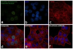
- Immunofluorescence analysis of Phospho-p53 (Ser15) was performed using 70% confluent log phase HCT116 cells treated with etoposide. The cells were fixed with 4% paraformaldehyde for 10 minutes, permeabilized with 0.1% Triton™ X-100 for 10 minutes, and blocked with 1% BSA for 1 hour at room temperature. The cells were labeled with Phospho-p53 Monoclonal Antibody(C3810) (Product # MA5-15229) at 1:100 dilution in 0.1% BSA, incubated overnight at 4 degree Celsius and then labeled with Goat anti-Mouse IgG (H+L) Superclonal™ Secondary Antibody, Alexa Fluor® 488 conjugate (Product # A28175) at a dilution of 1:2000 for 45 minutes at room temperature (Panel a: green). Nuclei (Panel b: blue) were stained with SlowFade® Gold Antifade Mountant with DAPI (Product # S36938). F-actin (Panel c: red) was stained with Rhodamine Phalloidin (Product # R415, 1:300). Panel d represents the merged image showing nuclear localization. Panel e represents the untreated cells with relatively lower expression of Phospho-p53(Ser15). Panel f shows control cells with no primary antibody to assess background. The images were captured at 60X magnification.

- Submitted by
- Invitrogen Antibodies (provider)
- Main image
- Experimental details
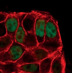
- Immunofluorescent analysis of Phospho-p53 pSer15 in HT-29 cells using a Phospho-p53 pSer15 monoclonal antibody (Product # MA5-15229) (green). Actin filaments are labeled with a fluorescent red phalloidin.

- Submitted by
- Invitrogen Antibodies (provider)
- Main image
- Experimental details
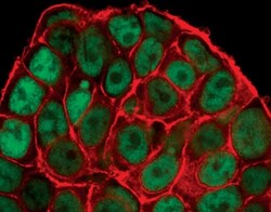
- Immunofluorescent analysis of Phospho-p53 pSer15 in HT-29 cells, UV-treated, using a Phospho-p53 pSer15 monoclonal antibody (Product # MA5-15229) (green). Actin filaments are labeled with a fluorescent red phalloidin.

Supportive validation
- Submitted by
- Invitrogen Antibodies (provider)
- Main image

- Experimental details
- Flow cytometric analysis of Phospho-p53 pSer15 in untreated (blue) or UV-treated (green) HT-29 cells using a Phospho-p53 pSer15 monoclonal antibody (Product # MA5-15229) compared to a nonspecific negative control antibody (red).

Supportive validation
- Submitted by
- Invitrogen Antibodies (provider)
- Main image

- Experimental details
- Figure 7 Phosphorylated p53 (p-p53), total p-53 (t-p53), and beta-actin expression as determined by Western blot ( a ) and phosphorylated p53 (p-p53) determined by confocal microscopy ( d ) in DLD-1 and HT-29 cells treated with 5-FU (5-FU 50 uM), MM-129 (MM 1 uM, 10 uM, 100 uM), and their combination (MM 10 uM + 5-FU 50 uM) for 24 h. The samples used for electrophoresis consisted of 20 ug of protein from 6 pooled cell extracts. The samples used for electrophoresis consisted of 20 ug of protein from 6 pooled cell extracts from independent experiments ( n = 6). Band staining was quantified by densitometry ( b , c ). The corresponding uncropped blots are shown in Supplementary Figures S7a-c and S8a-c . Cells were incubated with mouse monoclonal antibody against phospho p53 and FITC-conjugated secondary goat polyclonal antibody against mouse (red label). The nuclei were stained with Hoechst 33342 (blue label) ( d ). The results are presented as means +- SDs. * p < 0.05, ** p < 0.01, *** p < 0.001 vs. CON, ^^^ p < 0.001 vs. 5-FU, ### p < 0.001 vs. MM-129 at dose 10 uM.
