Explore
 Validate
 Learn
Learn Western blot
 Flow cytometry
Antibody data
- Antibody Data
- Antigen structure
- References [14]
- Comments [0]
- Validations
- Western blot [2]
- Immunocytochemistry [1]
- Immunohistochemistry [1]
Submit
Validation data
Reference
Comment
Report error
- Product number
- MA5-16387 - Provider product page

- Provider
- Invitrogen Antibodies
- Product name
- p53 Monoclonal Antibody (SP5)
- Antibody type
- Monoclonal
- Antigen
- Recombinant full-length protein
- Description
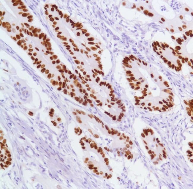
- Heat-mediated antigen retrieval is recommended prior to staining, using a 10mM citrate buffer, pH 6.0, for 10 minutes followed by cooling at room temperature for 20 min. Following antigen retrieval, incubate samples with primary antibody for 30 min at room temperature. A suggested positive control is colon carcinoma.
- Reactivity
- Human
- Host
- Rabbit
- Isotype
- IgG
- Antibody clone number
- SP5
- Vial size
- 500 µL
- Concentration
- 0.004 mg/mL
- Storage
- Store at 4°C short term. For long term storage, store at -20°C, avoiding freeze/thaw cycles.
Submitted references Simple Prediction Model of Axillary Lymph Node Positivity After Analyzing Molecular and Clinical Factors in Early Breast Cancer.
Quantitative apparent diffusion coefficient measurements obtained by 3-Tesla MRI are correlated with biomarkers of bladder cancer proliferative activity.
Evaluation of correlation of cell cycle proteins and Ki-67 interaction in paranasal sinus inverted papilloma prognosis and squamous cell carcinoma transformation.
Tissue biomarkers in prognostication of serous ovarian cancer following neoadjuvant chemotherapy.
Quantitative analysis of γ-H2AX and p53 nuclear expression levels in ovarian and fallopian tube epithelium from risk-reducing salpingo-oophorectomies in BRCA1 and BRCA2 mutation carriers.
Immunohistochemistry with apoptotic-antiapoptotic proteins (p53, p21, bax, bcl-2), c-kit, telomerase, and metallothionein as a diagnostic aid in benign, borderline, and malignant serous and mucinous ovarian tumors.
Genetic characterization of mesenchymal, clear cell, and dedifferentiated chondrosarcoma.
Proliferation and maturation of microvessels in arteriovenous malformations--expression patterns of angiogenic and cell cycle-dependent factors.
Evaluation of immunohistochemical expression of p53, p21, p27, cyclin D1, and Ki67 in oral and oropharyngeal squamous cell carcinoma.
Correlation of immunohistochemical expression of p53 with unamplified chromosome 17 polysomy in invasive breast carcinoma.
Extent and patterns of MGMT promoter methylation in glioblastoma- and respective glioblastoma-derived spheres.
Osteosarcoma arising in a long-standing uterine leiomyoma: a case report and literature review.
Recurrence of benign meningiomas: predictive value of proliferative index, BCL2, p53, hormonal receptors and HER2 expression.
Expression of senescence-related genes in human corneal endothelial cells.
Chung MJ, Lee JH, Kim SH, Suh YJ, Choi HJ
Medicine 2016 May;95(20):e3689
Medicine 2016 May;95(20):e3689
Quantitative apparent diffusion coefficient measurements obtained by 3-Tesla MRI are correlated with biomarkers of bladder cancer proliferative activity.
Sevcenco S, Haitel A, Ponhold L, Susani M, Fajkovic H, Shariat SF, Hiess M, Spick C, Szarvas T, Baltzer PA
PloS one 2014;9(9):e106866
PloS one 2014;9(9):e106866
Evaluation of correlation of cell cycle proteins and Ki-67 interaction in paranasal sinus inverted papilloma prognosis and squamous cell carcinoma transformation.
Tsou YA, Huang HJ, Wang TC, Tai CJ, Chen CM, Chen CY
BioMed research international 2014;2014:634945
BioMed research international 2014;2014:634945
Tissue biomarkers in prognostication of serous ovarian cancer following neoadjuvant chemotherapy.
Khandakar B, Mathur SR, Kumar L, Kumar S, Datta Gupta S, Iyer VK, Kalaivani M
BioMed research international 2014;2014:401245
BioMed research international 2014;2014:401245
Quantitative analysis of γ-H2AX and p53 nuclear expression levels in ovarian and fallopian tube epithelium from risk-reducing salpingo-oophorectomies in BRCA1 and BRCA2 mutation carriers.
Staff S, Tolonen T, Laasanen SL, Mecklin JP, Isola J, Mäenpää J
International journal of gynecological pathology : official journal of the International Society of Gynecological Pathologists 2014 May;33(3):309-16
International journal of gynecological pathology : official journal of the International Society of Gynecological Pathologists 2014 May;33(3):309-16
Immunohistochemistry with apoptotic-antiapoptotic proteins (p53, p21, bax, bcl-2), c-kit, telomerase, and metallothionein as a diagnostic aid in benign, borderline, and malignant serous and mucinous ovarian tumors.
Ozer H, Yenicesu G, Arici S, Cetin M, Tuncer E, Cetin A
Diagnostic pathology 2012 Sep 20;7:124
Diagnostic pathology 2012 Sep 20;7:124
Genetic characterization of mesenchymal, clear cell, and dedifferentiated chondrosarcoma.
Meijer D, de Jong D, Pansuriya TC, van den Akker BE, Picci P, Szuhai K, Bovée JV
Genes, chromosomes & cancer 2012 Oct;51(10):899-909
Genes, chromosomes & cancer 2012 Oct;51(10):899-909
Proliferation and maturation of microvessels in arteriovenous malformations--expression patterns of angiogenic and cell cycle-dependent factors.
Meijer-Jorna LB, van der Loos CM, Teeling P, de Boer OJ, Florquin S, van der Horst CM, van der Wal AC
Journal of cutaneous pathology 2012 Jun;39(6):610-20
Journal of cutaneous pathology 2012 Jun;39(6):610-20
Evaluation of immunohistochemical expression of p53, p21, p27, cyclin D1, and Ki67 in oral and oropharyngeal squamous cell carcinoma.
Perisanidis C, Perisanidis B, Wrba F, Brandstetter A, El Gazzar S, Papadogeorgakis N, Seemann R, Ewers R, Kyzas PA, Filipits M
Journal of oral pathology & medicine : official publication of the International Association of Oral Pathologists and the American Academy of Oral Pathology 2012 Jan;41(1):40-6
Journal of oral pathology & medicine : official publication of the International Association of Oral Pathologists and the American Academy of Oral Pathology 2012 Jan;41(1):40-6
Correlation of immunohistochemical expression of p53 with unamplified chromosome 17 polysomy in invasive breast carcinoma.
Krishnamurti U, Zarineh A, Atem FD, Silverman JF
Applied immunohistochemistry & molecular morphology : AIMM 2011 Jan;19(1):28-32
Applied immunohistochemistry & molecular morphology : AIMM 2011 Jan;19(1):28-32
Extent and patterns of MGMT promoter methylation in glioblastoma- and respective glioblastoma-derived spheres.
Sciuscio D, Diserens AC, van Dommelen K, Martinet D, Jones G, Janzer RC, Pollo C, Hamou MF, Kaina B, Stupp R, Levivier M, Hegi ME
Clinical cancer research : an official journal of the American Association for Cancer Research 2011 Jan 15;17(2):255-66
Clinical cancer research : an official journal of the American Association for Cancer Research 2011 Jan 15;17(2):255-66
Osteosarcoma arising in a long-standing uterine leiomyoma: a case report and literature review.
Wang RC, Wen MC, Wang J, Ho SC, Jan YJ
International journal of surgical pathology 2011 Feb;19(1):99-103
International journal of surgical pathology 2011 Feb;19(1):99-103
Recurrence of benign meningiomas: predictive value of proliferative index, BCL2, p53, hormonal receptors and HER2 expression.
Abdelzaher E, El-Gendi SM, Yehya A, Gowil AG
British journal of neurosurgery 2011 Dec;25(6):707-13
British journal of neurosurgery 2011 Dec;25(6):707-13
Expression of senescence-related genes in human corneal endothelial cells.
Song Z, Wang Y, Xie L, Zang X, Yin H
Molecular vision 2008 Jan 29;14:161-70
Molecular vision 2008 Jan 29;14:161-70
No comments: Submit comment
Supportive validation
- Submitted by
- Invitrogen Antibodies (provider)
- Main image
- Experimental details
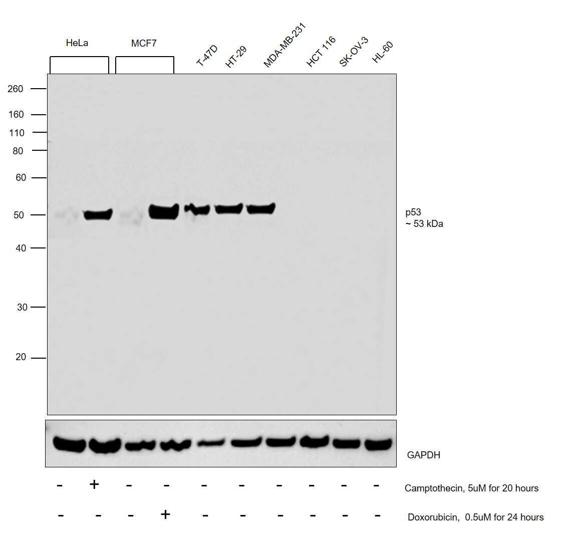
- Western blot was performed using Anti-p53 Monoclonal Antibody (SP5) (Product # MA5-16387) and a 53 kDa band corresponding to Cellular tumor antigen p53 was observed across cell lines except HCT 116, SK-OV-3 and HL-60 which are reported to be negative, also it was observed to be induced upon Camptothecin and Doxorubicin treatment in HeLa and MCF7 respectively. Modified whole cell extracts (1%SDS) (30 µg lysate) of HeLa (Lane 1), HeLa treated with Camptothecin (5uM for 20 hours) (Lane 2), MCF7 (Lane 3), MCF7 treated with Doxorubicin (0.5uM for 24 hours) (Lane 4), T-47D (Lane 5), HT-29 (Lane 6), MDA-MB-231 (Lane 7), HCT 116 (Lane 8), SK-O-V3 (Lane 9) and HL-60 (Lane 10) were electrophoresed using NuPAGE™ 10% Bis-Tris Protein Gel (Product # NP0302BOX). Resolved proteins were then transferred onto a Nitrocellulose membrane (Product # IB23001) by iBlot® 2 Dry Blotting System (Product # IB21001). The blot was probed with the primary antibody (1:1000 dilution) and detected by chemiluminescence with Goat anti-Rabbit IgG (H+L) Superclonal™ Recombinant Secondary Antibody, HRP (Product # A27036,1:4000 dilution) using the iBright FL 1000 (Product # A32752). Chemiluminescent detection was performed using Novex® ECL Chemiluminescent Substrate Reagent Kit (Product # WP20005).
- Submitted by
- Invitrogen Antibodies (provider)
- Main image

- Experimental details
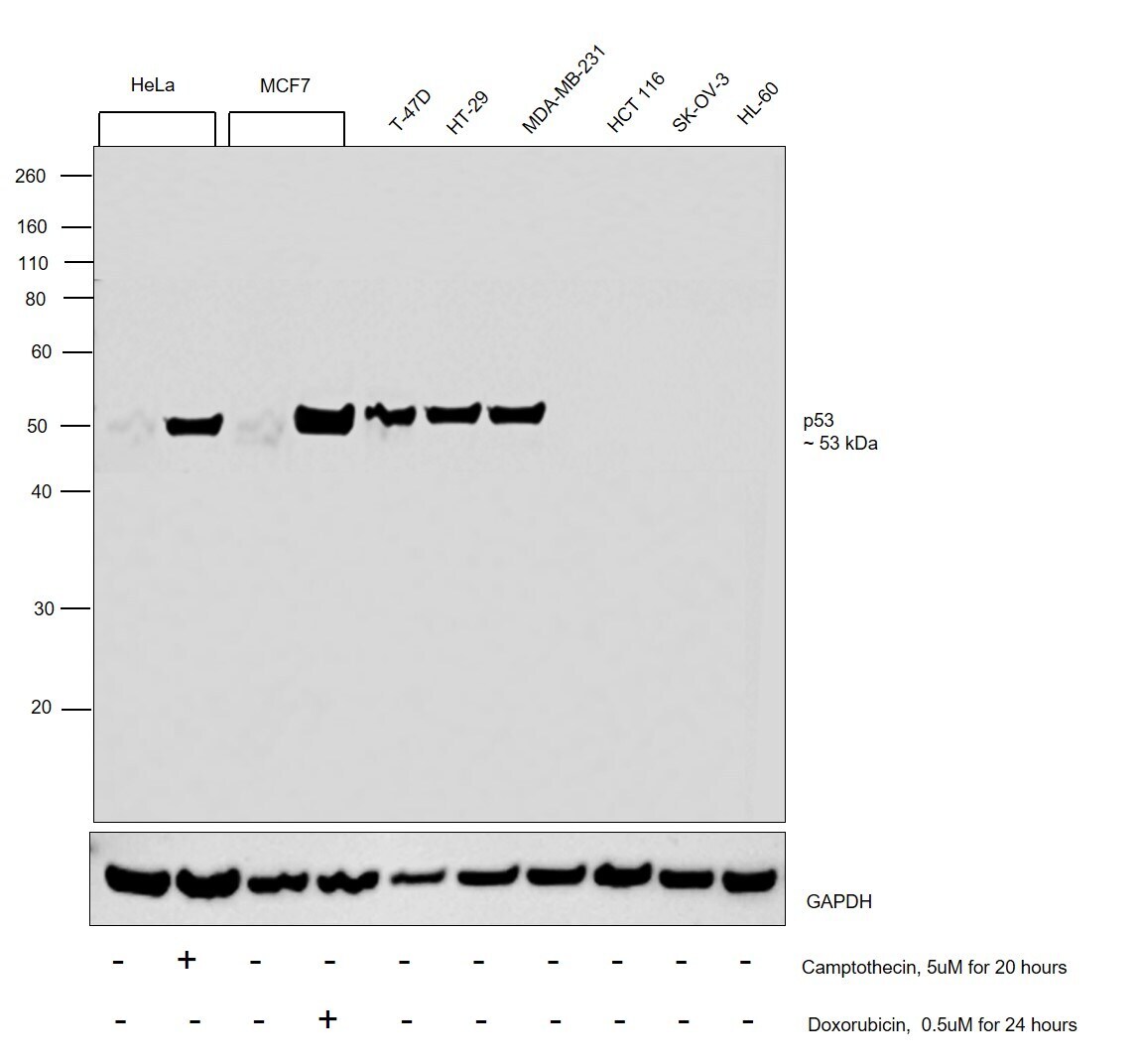
- Western blot was performed using Anti-p53 Monoclonal Antibody (SP5) (Product # MA5-16387) and a 53 kDa band corresponding to Cellular tumor antigen p53 was observed across cell lines except HCT 116, SK-OV-3 and HL-60 which are reported to be negative, also it was observed to be induced upon Camptothecin and Doxorubicin treatment in HeLa and MCF7 respectively. Modified whole cell extracts (1%SDS) (30 µg lysate) of HeLa (Lane 1), HeLa treated with Camptothecin (5uM for 20 hours) (Lane 2), MCF7 (Lane 3), MCF7 treated with Doxorubicin (0.5uM for 24 hours) (Lane 4), T-47D (Lane 5), HT-29 (Lane 6), MDA-MB-231 (Lane 7), HCT 116 (Lane 8), SK-O-V3 (Lane 9) and HL-60 (Lane 10) were electrophoresed using NuPAGE™ 10% Bis-Tris Protein Gel (Product # NP0302BOX). Resolved proteins were then transferred onto a Nitrocellulose membrane (Product # IB23001) by iBlot® 2 Dry Blotting System (Product # IB21001). The blot was probed with the primary antibody (1:1000 dilution) and detected by chemiluminescence with Goat anti-Rabbit IgG (H+L) Superclonal™ Recombinant Secondary Antibody, HRP (Product # A27036,1:4000 dilution) using the iBright FL 1000 (Product # A32752). Chemiluminescent detection was performed using Novex® ECL Chemiluminescent Substrate Reagent Kit (Product # WP20005).
Supportive validation
- Submitted by
- Invitrogen Antibodies (provider)
- Main image
- Experimental details
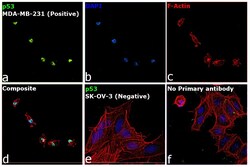
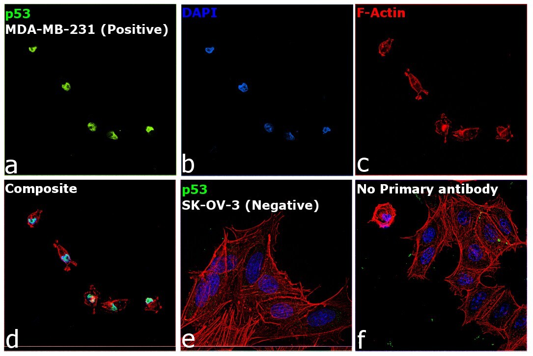
- Immunofluorescence analysis of Cellular tumor antigen p53 was performed using 70% confluent log phase MDA-MB-231 cells. The cells were fixed with 4% paraformaldehyde for 10 minutes, permeabilized with 0.1% Triton™ X-100 for 15 minutes, and blocked with 2% BSA for 45 minutes at room temperature. The cells were labeled with p53 Monoclonal Antibody (SP5) (Product # MA5-16387) at 1:100 dilution in 0.1% BSA, incubated at 4 degree celsius overnight and then labeled with Donkey anti-Rabbit IgG (H+L) Highly Cross-Adsorbed Secondary Antibody, Alexa Fluor Plus 488 (Product # A32790), (1:2000 dilution), for 45 minutes at room temperature (Panel a: Green). Nuclei (Panel b: Blue) were stained with ProLong™ Diamond Antifade Mountant with DAPI (Product # P36962). F-actin (Panel c: Red) was stained with Rhodamine Phalloidin (Product # R415, 1:300). Panel d represents the merged image showing Nuclear localization. Panel e represents SK-OV-3 cells having no expression of p53. Panel f represents control cells with no primary antibody to assess background. The images were captured at 60X magnification.
Supportive validation
- Submitted by
- Invitrogen Antibodies (provider)
- Main image

- Experimental details
- Immunohistochemical analysis of p53 using anti-p53 Monoclonal Antibody (Product # MA5-16387) in Colon Carcinoma Cancer Tissue. The recommened dilution for this antibody in immunohistochemistry applications is 1:100.