 Explore
 Validate
 Learn
Learn Western blot
Antibody data
- Antibody Data
- Antigen structure
- References [1]
- Comments [0]
- Validations
- Western blot [1]
- Immunohistochemistry [1]
- Other assay [3]
Submit
Validation data
Reference
Comment
Report error
- Product number
- PA5-12660 - Provider product page

- Provider
- Invitrogen Antibodies
- Product name
- Phospho-p53 (Thr18) Polyclonal Antibody
- Antibody type
- Polyclonal
- Antigen
- Synthetic peptide
- Description
- This antibody is predicted to react with porcine, non-human primate and rabbit based on sequence homology.
- Reactivity
- Human, Mouse
- Host
- Rabbit
- Isotype
- IgG
- Vial size
- 400 µL
- Concentration
- 0.5 mg/mL
- Storage
- -20° C, Avoid Freeze/Thaw Cycles
Submitted references Phosphorylation of vaccinia-related kinase 1 at threonine 386 transduces glucose stress signal in human liver cells.
Yokobori K, Miyauchi Y, Williams JG, Negishi M
Bioscience reports 2020 Apr 30;40(4)
Bioscience reports 2020 Apr 30;40(4)
No comments: Submit comment
Supportive validation
- Submitted by
- Invitrogen Antibodies (provider)
- Main image

- Experimental details
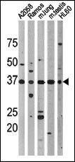
- Western blot analysis of, from left to right, A2058, Ramos, mouse lung, mouse testis, and HL-60 cell lysates using a Phospho-p53 pThr18 polyclonal antibody (Product # PA5-12660).
Supportive validation
- Submitted by
- Invitrogen Antibodies (provider)
- Main image

- Experimental details
- Immunohistochemical analysis of formalin-fixed, paraffin-embedded human cancer tissue using a Phospho-p53 pThr18 polyclonal antibody (Product # PA5-12660), followed by HRP-conjugated secondary antibody and AEC staining.

Supportive validation
- Submitted by
- Invitrogen Antibodies (provider)
- Main image

- Experimental details
- Figure 1 VRK1 activation by glucose stimulation Huh-7 cells were transfected with siVRK1 or control for 24 h in middle (100 mg/dl) glucose and, subsequently, in low (40 mg/dl) or high (140 mg/dl) glucose for 3 h. Whole extracts of these cells were prepared using urea buffer. c-Jun and p53 phosphorylation were analyzed by Western blotting using an anti-P-Ser63 c-Jun antibody and an anti-P-Thr18 p53 antibody, respectively. Protein expression levels of c-Jun, p53 or VRK1 were determined by Western blotting assay. Expression levels of beta-actin were used for loading control.

- Submitted by
- Invitrogen Antibodies (provider)
- Main image

- Experimental details
- Figure 5 Regulation of VRK1 activity by phosphorylation Huh-7 cells were expressed with FLAG-tagged VRK1-WT, T386A, T386D, or mock for 24 h in high (140 mg/dl) glucose. Whole extracts of these cells were prepared using urea buffer. c-Jun and p53 phosphorylation were analyzed by Western blotting using an anti-P-Ser63 c-Jun antibody and an anti-P-Thr18 p53 antibody, respectively. Protein expression levels of c-Jun, p53 or FLAG-tagged VRK1 were determined by Western blotting. Expression levels of beta-actin were used for loading control.

- Submitted by
- Invitrogen Antibodies (provider)
- Main image
- Experimental details
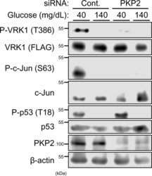
- Figure 7 Regulation of VRK1 activity by PKP2 After transfection with siPKP2 or control for 24 h, Huh-7 cells were expressed with FLAG-tagged VRK1 for 24 h in middle (100 mg/dl) glucose and, subsequently, in low (40 mg/dl) or high (140 mg/dl) glucose for 3 h. Whole extracts of these cells were prepared using IP buffer. Phosphorylated FLAG-tagged VRK1 was immunoprecipitated by anti-phospho VRK1 antibodies conjugated with Dynabeads Protein G. The resultant precipitates and whole cell extracts were analyzed by Western blotting for phospho-VRK1 and total FLAG-tagged VRK1, respectively, using an anti-FLAG HRP conjugated antibody. c-Jun and p53 phosphorylation were analyzed by Western blotting using an anti-P-Ser63 c-Jun antibody and an anti-P-Thr18 p53 antibody, respectively. Protein expression levels of c-Jun, p53 or PKP2 were determined by Western blotting. Expression levels of beta-actin were used for loading control.
