 Explore
 Validate
 Learn
Learn Western blot
Antibody data
- Antibody Data
- Antigen structure
- References [68]
- Comments [0]
- Validations
- Western blot [4]
- Immunocytochemistry [3]
- Immunoprecipitation [2]
- Immunohistochemistry [1]
- Other assay [9]
Submit
Validation data
Reference
Comment
Report error
- Product number
- MA5-12571 - Provider product page

- Provider
- Invitrogen Antibodies
- Product name
- p53 Monoclonal Antibody (DO-1)
- Antibody type
- Monoclonal
- Antigen
- Recombinant full-length protein
- Description
- MA5-12571 targets p53 in immunohistochemistry (paraffin) and Western blot applications and shows reactivity with Human samples.
- Antibody clone number
- DO-1
- Concentration
- 0.2 mg/mL
Submitted references AHNAK controls 53BP1-mediated p53 response by restraining 53BP1 oligomerization and phase separation.
TIRR inhibits the 53BP1-p53 complex to alter cell-fate programs.
ABHD5 suppresses cancer cell anabolism through lipolysis-dependent activation of the AMPK/mTORC1 pathway.
Increased p53 signaling impairs neural differentiation in HUWE1-promoted intellectual disabilities.
Targeting mutant p53-expressing tumours with a T cell receptor-like antibody specific for a wild-type antigen.
Essential Gene Profiles for Human Pluripotent Stem Cells Identify Uncharacterized Genes and Substrate Dependencies.
Tyrosine kinase c-Abl couples RNA polymerase II transcription to DNA double-strand breaks.
p53 Regulates the Expression of LRP1 and Apoptosis through a Stress Intensity-Dependent MicroRNA Feedback Loop.
Metformin produces growth inhibitory effects in combination with nutlin-3a on malignant mesothelioma through a cross-talk between mTOR and p53 pathways.
High-Risk HPV, Biomarkers, and Outcome in Matched Cohorts of Head and Neck Cancer Patients Positive and Negative for HIV.
Preclinical Efficacy of the MDM2 Inhibitor RG7112 in MDM2-Amplified and TP53 Wild-type Glioblastomas.
MAP3K8/TPL-2/COT is a potential predictive marker for MEK inhibitor treatment in high-grade serous ovarian carcinomas.
The MDM2 RING domain and central acidic domain play distinct roles in MDM2 protein homodimerization and MDM2-MDMX protein heterodimerization.
Acquisition of Portal Venous Circulating Tumor Cells From Patients With Pancreaticobiliary Cancers by Endoscopic Ultrasound.
FATS is an E2-independent ubiquitin ligase that stabilizes p53 and promotes its activation in response to DNA damage.
Matricellular protein CCN1 (CYR61) expression is associated with high-grade ductal carcinoma in situ.
Enhanced anticancer activity and circumvention of resistance mechanisms by novel polymeric/ phospholipidic nanocarriers of doxorubicin.
Enhanced anticancer activity and circumvention of resistance mechanisms by novel polymeric/ phospholipidic nanocarriers of doxorubicin.
Biomarkers in advanced larynx cancer.
Invasive ductular carcinoma in 2 rhesus macaques (Macaca mulatta).
Head and neck squamous cell carcinoma in pregnant women.
A non-catalytic role of DNA polymerase η in recruiting Rad18 and promoting PCNA monoubiquitination at stalled replication forks.
Merkel cell polyomavirus large T antigen has growth-promoting and inhibitory activities.
Influence of extracellular pH on the cytotoxicity, cellular accumulation, and DNA interaction of novel pH-sensitive 2-aminoalcoholatoplatinum(II) complexes.
Destruxins: fungal-derived cyclohexadepsipeptides with multifaceted anticancer and antiangiogenic activities.
Mdm2 RING mutation enhances p53 transcriptional activity and p53-p300 interaction.
Trabectedin has promising antineoplastic activity in high-grade meningioma.
Resistance to butyrate impairs bile acid-induced apoptosis in human colon adenocarcinoma cells via up-regulation of Bcl-2 and inactivation of Bax.
Deoxycholic and chenodeoxycholic bile acids induce apoptosis via oxidative stress in human colon adenocarcinoma cells.
p53-Inducible DHRS3 is an endoplasmic reticulum protein associated with lipid droplet accumulation.
An ARF-independent c-MYC-activated tumor suppression pathway mediated by ribosomal protein-Mdm2 Interaction.
Interaction with Sug1 enables Ipaf ubiquitination leading to caspase 8 activation and cell death.
Response of experimental malignant melanoma models to the pan-Aurora kinase inhibitor VE-465.
Mitochondrial Hep27 is a c-Myb target gene that inhibits Mdm2 and stabilizes p53.
Regulation of myeloid leukaemia by the cell-fate determinant Musashi.
Repression of the miR-17-92 cluster by p53 has an important function in hypoxia-induced apoptosis.
Differences in the nemosis response of normal and cancer-associated fibroblasts from patients with oral squamous cell carcinoma.
Establishment of ponasterone A-inducible the wild-type p53 protein-expressing clones from HSC-1 cells, cell growth suppression by p53 expression and the suppression mechanism.
Oncolytic adenoviral vectors which employ the survivin promoter induce glioma oncolysis via a process of beclin-dependent autophagy.
Simultaneous blockade of the epidermal growth factor receptor/mammalian target of rapamycin pathway by epidermal growth factor receptor inhibitors and rapamycin results in reduced cell growth and survival in biliary tract cancer cells.
p53 Oligomerization is essential for its C-terminal lysine acetylation.
Her-2/neu, p-53, and their coexpression in osteosarcoma.
Combination of adenoviral virotherapy and temozolomide chemotherapy eradicates malignant glioma through autophagic and apoptotic cell death in vivo.
The transduction of His-TAT-p53 fusion protein into the human osteogenic sarcoma cell line (Saos-2) and its influence on cell cycle arrest and apoptosis.
Mitochondrial p32 is a critical mediator of ARF-induced apoptosis.
Experimental validation for quantitative protein network models.
p53 signaling in response to increased DNA damage sensitizes AML1-ETO cells to stress-induced death.
p53 gene mutational rate, Gleason score, and BK virus infection in prostate adenocarcinoma: Is there a correlation?
Expression of p53 and Bcl-xL as predictive markers for larynx preservation in advanced laryngeal cancer.
Targeting apoptosis to overcome cisplatin resistance: a translational study in head and neck cancer.
p53 and mdm2 expression in colorectal carcinoma: a correlative analysis with clinical staging and histological parameters.
PARC and CUL7 form atypical cullin RING ligase complexes.
Antitumor activity of CTFB, a novel anticancer agent, is associated with the down-regulation of nuclear factor-kappaB expression and proteasome activation in head and neck squamous carcinoma cell lines.
The ubiquitin-proteasome system regulates p53-mediated transcription at p21waf1 promoter.
p53-Dependent but ATM-independent inhibition of DNA synthesis and G2 arrest in cadmium-treated human fibroblasts.
Human cytomegalovirus disrupts both ataxia telangiectasia mutated protein (ATM)- and ATM-Rad3-related kinase-mediated DNA damage responses during lytic infection.
Adenovirus-mediated p53 gene transfer sensitizes hepatocellular carcinoma cells to heavy-ion radiation.
A novel p53-binding domain in CUL7.
Targeting of p300/CREB binding protein coactivators by simian virus 40 is mediated through p53.
p53 suppression of arsenite-induced mitotic catastrophe is mediated by p21CIP1/WAF1.
Telomerase-immortalized human fibroblasts retain UV-induced mutagenesis and p53-mediated DNA damage responses.
Simian virus 40 large T antigen's association with the CUL7 SCF complex contributes to cellular transformation.
Tumor suppressor HIC1 directly regulates SIRT1 to modulate p53-dependent DNA-damage responses.
Detection and expression of human BK virus sequences in neoplastic prostate tissues.
Stabilization of alanine substituted p53 protein at Ser15, Thr18, and Ser20 in response to ionizing radiation.
Increased apoptosis in human osteoarthritic cartilage corresponds to reduced cell density and expression of caspase-3.
Photodynamic therapy sensitivity is not altered in human tumor cells after abrogation of p53 function.
Loss of butyrate-induced apoptosis in human hepatoma cell lines HCC-M and HCC-T having substantial Bcl-2 expression.
Ghodke I, Remisova M, Furst A, Kilic S, Reina-San-Martin B, Poetsch AR, Altmeyer M, Soutoglou E
Molecular cell 2021 Jun 17;81(12):2596-2610.e7
Molecular cell 2021 Jun 17;81(12):2596-2610.e7
TIRR inhibits the 53BP1-p53 complex to alter cell-fate programs.
Parnandi N, Rendo V, Cui G, Botuyan MV, Remisova M, Nguyen H, Drané P, Beroukhim R, Altmeyer M, Mer G, Chowdhury D
Molecular cell 2021 Jun 17;81(12):2583-2595.e6
Molecular cell 2021 Jun 17;81(12):2583-2595.e6
ABHD5 suppresses cancer cell anabolism through lipolysis-dependent activation of the AMPK/mTORC1 pathway.
Chen G, Zhou G, Lotvola A, Granneman JG, Wang J
The Journal of biological chemistry 2021 Jan-Jun;296:100104
The Journal of biological chemistry 2021 Jan-Jun;296:100104
Increased p53 signaling impairs neural differentiation in HUWE1-promoted intellectual disabilities.
Aprigliano R, Aksu ME, Bradamante S, Mihaljevic B, Wang W, Rian K, Montaldo NP, Grooms KM, Fordyce Martin SL, Bordin DL, Bosshard M, Peng Y, Alexov E, Skinner C, Liabakk NB, Sullivan GJ, Bjørås M, Schwartz CE, van Loon B
Cell reports. Medicine 2021 Apr 20;2(4):100240
Cell reports. Medicine 2021 Apr 20;2(4):100240
Targeting mutant p53-expressing tumours with a T cell receptor-like antibody specific for a wild-type antigen.
Low L, Goh A, Koh J, Lim S, Wang CI
Nature communications 2019 Nov 26;10(1):5382
Nature communications 2019 Nov 26;10(1):5382
Essential Gene Profiles for Human Pluripotent Stem Cells Identify Uncharacterized Genes and Substrate Dependencies.
Mair B, Tomic J, Masud SN, Tonge P, Weiss A, Usaj M, Tong AHY, Kwan JJ, Brown KR, Titus E, Atkins M, Chan KSK, Munsie L, Habsid A, Han H, Kennedy M, Cohen B, Keller G, Moffat J
Cell reports 2019 Apr 9;27(2):599-615.e12
Cell reports 2019 Apr 9;27(2):599-615.e12
Tyrosine kinase c-Abl couples RNA polymerase II transcription to DNA double-strand breaks.
Burger K, Schlackow M, Gullerova M
Nucleic acids research 2019 Apr 23;47(7):3467-3484
Nucleic acids research 2019 Apr 23;47(7):3467-3484
p53 Regulates the Expression of LRP1 and Apoptosis through a Stress Intensity-Dependent MicroRNA Feedback Loop.
Leslie PL, Franklin DA, Liu Y, Zhang Y
Cell reports 2018 Aug 7;24(6):1484-1495
Cell reports 2018 Aug 7;24(6):1484-1495
Metformin produces growth inhibitory effects in combination with nutlin-3a on malignant mesothelioma through a cross-talk between mTOR and p53 pathways.
Shimazu K, Tada Y, Morinaga T, Shingyoji M, Sekine I, Shimada H, Hiroshima K, Namiki T, Tatsumi K, Tagawa M
BMC cancer 2017 May 2;17(1):309
BMC cancer 2017 May 2;17(1):309
High-Risk HPV, Biomarkers, and Outcome in Matched Cohorts of Head and Neck Cancer Patients Positive and Negative for HIV.
Walline HM, Carey TE, Goudsmit CM, Bellile EL, D'Souza G, Peterson LA, McHugh JB, Pai SI, Lee JJ, Shin DM, Ferris RL, HNC SPORE HIV supplement consortium
Molecular cancer research : MCR 2017 Feb;15(2):179-188
Molecular cancer research : MCR 2017 Feb;15(2):179-188
Preclinical Efficacy of the MDM2 Inhibitor RG7112 in MDM2-Amplified and TP53 Wild-type Glioblastomas.
Verreault M, Schmitt C, Goldwirt L, Pelton K, Haidar S, Levasseur C, Guehennec J, Knoff D, Labussière M, Marie Y, Ligon AH, Mokhtari K, Hoang-Xuan K, Sanson M, Alexander BM, Wen PY, Delattre JY, Ligon KL, Idbaih A
Clinical cancer research : an official journal of the American Association for Cancer Research 2016 Mar 1;22(5):1185-96
Clinical cancer research : an official journal of the American Association for Cancer Research 2016 Mar 1;22(5):1185-96
MAP3K8/TPL-2/COT is a potential predictive marker for MEK inhibitor treatment in high-grade serous ovarian carcinomas.
Gruosso T, Garnier C, Abelanet S, Kieffer Y, Lemesre V, Bellanger D, Bieche I, Marangoni E, Sastre-Garau X, Mieulet V, Mechta-Grigoriou F
Nature communications 2015 Oct 12;6:8583
Nature communications 2015 Oct 12;6:8583
The MDM2 RING domain and central acidic domain play distinct roles in MDM2 protein homodimerization and MDM2-MDMX protein heterodimerization.
Leslie PL, Ke H, Zhang Y
The Journal of biological chemistry 2015 May 15;290(20):12941-50
The Journal of biological chemistry 2015 May 15;290(20):12941-50
Acquisition of Portal Venous Circulating Tumor Cells From Patients With Pancreaticobiliary Cancers by Endoscopic Ultrasound.
Catenacci DV, Chapman CG, Xu P, Koons A, Konda VJ, Siddiqui UD, Waxman I
Gastroenterology 2015 Dec;149(7):1794-1803.e4
Gastroenterology 2015 Dec;149(7):1794-1803.e4
FATS is an E2-independent ubiquitin ligase that stabilizes p53 and promotes its activation in response to DNA damage.
Yan S, Qiu L, Ma K, Zhang X, Zhao Y, Zhang J, Li X, Hao X, Li Z
Oncogene 2014 Nov 20;33(47):5424-33
Oncogene 2014 Nov 20;33(47):5424-33
Matricellular protein CCN1 (CYR61) expression is associated with high-grade ductal carcinoma in situ.
Saglam O, Dai F, Husain S, Zhan Y, Toruner G, Haines GK 3rd
Human pathology 2014 Jun;45(6):1269-75
Human pathology 2014 Jun;45(6):1269-75
Enhanced anticancer activity and circumvention of resistance mechanisms by novel polymeric/ phospholipidic nanocarriers of doxorubicin.
Senkiv Y, Riabtseva A, Heffeter P, Boiko N, Kowol CR, Jungwith U, Shlyakhtina Y, Garasevych SG, Mitina N, Berger W, Zaichenko A, Stoika R
Journal of biomedical nanotechnology 2014 Jul;10(7):1369-81
Journal of biomedical nanotechnology 2014 Jul;10(7):1369-81
Biomarkers in advanced larynx cancer.
Bradford CR, Kumar B, Bellile E, Lee J, Taylor J, D'Silva N, Cordell K, Kleer C, Kupfer R, Kumar P, Urba S, Worden F, Eisbruch A, Wolf GT, Teknos TN, Prince ME, Chepeha DB, Hogikyan ND, Moyer JS, Carey TE
The Laryngoscope 2014 Jan;124(1):179-87
The Laryngoscope 2014 Jan;124(1):179-87
Invasive ductular carcinoma in 2 rhesus macaques (Macaca mulatta).
Beck AP, Brooks A, Zeiss CJ
Comparative medicine 2014 Aug;64(4):314-22
Comparative medicine 2014 Aug;64(4):314-22
Head and neck squamous cell carcinoma in pregnant women.
Eliassen AM, Hauff SJ, Tang AL, Thomas DH, McHugh JB, Walline HM, Stoerker J, Maxwell JH, Worden FP, Eisbruch A, Czerwinski MJ, Papagerakis SM, Chepeha DB, Bradford CR, Hanauer DA, Carey TE, Prince ME
Head & neck 2013 Mar;35(3):335-42
Head & neck 2013 Mar;35(3):335-42
A non-catalytic role of DNA polymerase η in recruiting Rad18 and promoting PCNA monoubiquitination at stalled replication forks.
Durando M, Tateishi S, Vaziri C
Nucleic acids research 2013 Mar 1;41(5):3079-93
Nucleic acids research 2013 Mar 1;41(5):3079-93
Merkel cell polyomavirus large T antigen has growth-promoting and inhibitory activities.
Cheng J, Rozenblatt-Rosen O, Paulson KG, Nghiem P, DeCaprio JA
Journal of virology 2013 Jun;87(11):6118-26
Journal of virology 2013 Jun;87(11):6118-26
Influence of extracellular pH on the cytotoxicity, cellular accumulation, and DNA interaction of novel pH-sensitive 2-aminoalcoholatoplatinum(II) complexes.
Valiahdi SM, Egger AE, Miklos W, Jungwirth U, Meelich K, Nock P, Berger W, Hartinger CG, Galanski MS, Jakupec MA, Keppler BK
Journal of biological inorganic chemistry : JBIC : a publication of the Society of Biological Inorganic Chemistry 2013 Feb;18(2):249-260
Journal of biological inorganic chemistry : JBIC : a publication of the Society of Biological Inorganic Chemistry 2013 Feb;18(2):249-260
Destruxins: fungal-derived cyclohexadepsipeptides with multifaceted anticancer and antiangiogenic activities.
Dornetshuber-Fleiss R, Heffeter P, Mohr T, Hazemi P, Kryeziu K, Seger C, Berger W, Lemmens-Gruber R
Biochemical pharmacology 2013 Aug 1;86(3):361-77
Biochemical pharmacology 2013 Aug 1;86(3):361-77
Mdm2 RING mutation enhances p53 transcriptional activity and p53-p300 interaction.
Clegg HV, Itahana Y, Itahana K, Ramalingam S, Zhang Y
PloS one 2012;7(5):e38212
PloS one 2012;7(5):e38212
Trabectedin has promising antineoplastic activity in high-grade meningioma.
Preusser M, Spiegl-Kreinecker S, Lötsch D, Wöhrer A, Schmook M, Dieckmann K, Saringer W, Marosi C, Berger W
Cancer 2012 Oct 15;118(20):5038-49
Cancer 2012 Oct 15;118(20):5038-49
Resistance to butyrate impairs bile acid-induced apoptosis in human colon adenocarcinoma cells via up-regulation of Bcl-2 and inactivation of Bax.
Barrasa JI, Santiago-Gómez A, Olmo N, Lizarbe MA, Turnay J
Biochimica et biophysica acta 2012 Dec;1823(12):2201-9
Biochimica et biophysica acta 2012 Dec;1823(12):2201-9
Deoxycholic and chenodeoxycholic bile acids induce apoptosis via oxidative stress in human colon adenocarcinoma cells.
Ignacio Barrasa J, Olmo N, Pérez-Ramos P, Santiago-Gómez A, Lecona E, Turnay J, Antonia Lizarbe M
Apoptosis : an international journal on programmed cell death 2011 Oct;16(10):1054-67
Apoptosis : an international journal on programmed cell death 2011 Oct;16(10):1054-67
p53-Inducible DHRS3 is an endoplasmic reticulum protein associated with lipid droplet accumulation.
Deisenroth C, Itahana Y, Tollini L, Jin A, Zhang Y
The Journal of biological chemistry 2011 Aug 12;286(32):28343-56
The Journal of biological chemistry 2011 Aug 12;286(32):28343-56
An ARF-independent c-MYC-activated tumor suppression pathway mediated by ribosomal protein-Mdm2 Interaction.
Macias E, Jin A, Deisenroth C, Bhat K, Mao H, Lindström MS, Zhang Y
Cancer cell 2010 Sep 14;18(3):231-43
Cancer cell 2010 Sep 14;18(3):231-43
Interaction with Sug1 enables Ipaf ubiquitination leading to caspase 8 activation and cell death.
Kumar Y, Radha V, Swarup G
The Biochemical journal 2010 Mar 15;427(1):91-104
The Biochemical journal 2010 Mar 15;427(1):91-104
Response of experimental malignant melanoma models to the pan-Aurora kinase inhibitor VE-465.
Pirker C, Lötsch D, Spiegl-Kreinecker S, Jantscher F, Sutterlüty H, Micksche M, Grusch M, Berger W
Experimental dermatology 2010 Dec;19(12):1040-7
Experimental dermatology 2010 Dec;19(12):1040-7
Mitochondrial Hep27 is a c-Myb target gene that inhibits Mdm2 and stabilizes p53.
Deisenroth C, Thorner AR, Enomoto T, Perou CM, Zhang Y
Molecular and cellular biology 2010 Aug;30(16):3981-93
Molecular and cellular biology 2010 Aug;30(16):3981-93
Regulation of myeloid leukaemia by the cell-fate determinant Musashi.
Ito T, Kwon HY, Zimdahl B, Congdon KL, Blum J, Lento WE, Zhao C, Lagoo A, Gerrard G, Foroni L, Goldman J, Goh H, Kim SH, Kim DW, Chuah C, Oehler VG, Radich JP, Jordan CT, Reya T
Nature 2010 Aug 5;466(7307):765-8
Nature 2010 Aug 5;466(7307):765-8
Repression of the miR-17-92 cluster by p53 has an important function in hypoxia-induced apoptosis.
Yan HL, Xue G, Mei Q, Wang YZ, Ding FX, Liu MF, Lu MH, Tang Y, Yu HY, Sun SH
The EMBO journal 2009 Sep 16;28(18):2719-32
The EMBO journal 2009 Sep 16;28(18):2719-32
Differences in the nemosis response of normal and cancer-associated fibroblasts from patients with oral squamous cell carcinoma.
Räsänen K, Virtanen I, Salmenperä P, Grenman R, Vaheri A
PloS one 2009 Sep 1;4(9):e6879
PloS one 2009 Sep 1;4(9):e6879
Establishment of ponasterone A-inducible the wild-type p53 protein-expressing clones from HSC-1 cells, cell growth suppression by p53 expression and the suppression mechanism.
Hori M, Suzuki K, Udono MU, Yamauchi M, Mine M, Watanabe M, Kondo S, Hozumi Y
Archives of dermatological research 2009 Oct;301(9):631-46
Archives of dermatological research 2009 Oct;301(9):631-46
Oncolytic adenoviral vectors which employ the survivin promoter induce glioma oncolysis via a process of beclin-dependent autophagy.
Ulasov IV, Tyler MA, Zhu ZB, Han Y, He TC, Lesniak MS
International journal of oncology 2009 Mar;34(3):729-42
International journal of oncology 2009 Mar;34(3):729-42
Simultaneous blockade of the epidermal growth factor receptor/mammalian target of rapamycin pathway by epidermal growth factor receptor inhibitors and rapamycin results in reduced cell growth and survival in biliary tract cancer cells.
Herberger B, Berger W, Puhalla H, Schmid K, Novak S, Brandstetter A, Pirker C, Gruenberger T, Filipits M
Molecular cancer therapeutics 2009 Jun;8(6):1547-56
Molecular cancer therapeutics 2009 Jun;8(6):1547-56
p53 Oligomerization is essential for its C-terminal lysine acetylation.
Itahana Y, Ke H, Zhang Y
The Journal of biological chemistry 2009 Feb 20;284(8):5158-64
The Journal of biological chemistry 2009 Feb 20;284(8):5158-64
Her-2/neu, p-53, and their coexpression in osteosarcoma.
Bakhshi S, Gupta A, Sharma MC, Khan SA, Rastogi S
Journal of pediatric hematology/oncology 2009 Apr;31(4):245-51
Journal of pediatric hematology/oncology 2009 Apr;31(4):245-51
Combination of adenoviral virotherapy and temozolomide chemotherapy eradicates malignant glioma through autophagic and apoptotic cell death in vivo.
Ulasov IV, Sonabend AM, Nandi S, Khramtsov A, Han Y, Lesniak MS
British journal of cancer 2009 Apr 7;100(7):1154-64
British journal of cancer 2009 Apr 7;100(7):1154-64
The transduction of His-TAT-p53 fusion protein into the human osteogenic sarcoma cell line (Saos-2) and its influence on cell cycle arrest and apoptosis.
Jiang L, Ma Y, Wang J, Tao X, Wei D
Molecular biology reports 2008 Mar;35(1):1-8
Molecular biology reports 2008 Mar;35(1):1-8
Mitochondrial p32 is a critical mediator of ARF-induced apoptosis.
Itahana K, Zhang Y
Cancer cell 2008 Jun;13(6):542-53
Cancer cell 2008 Jun;13(6):542-53
Experimental validation for quantitative protein network models.
Nishizuka S, Spurrier B
Current opinion in biotechnology 2008 Feb;19(1):41-9
Current opinion in biotechnology 2008 Feb;19(1):41-9
p53 signaling in response to increased DNA damage sensitizes AML1-ETO cells to stress-induced death.
Krejci O, Wunderlich M, Geiger H, Chou FS, Schleimer D, Jansen M, Andreassen PR, Mulloy JC
Blood 2008 Feb 15;111(4):2190-9
Blood 2008 Feb 15;111(4):2190-9
p53 gene mutational rate, Gleason score, and BK virus infection in prostate adenocarcinoma: Is there a correlation?
Russo G, Anzivino E, Fioriti D, Mischitelli M, Bellizzi A, Giordano A, Autran-Gomez A, Di Monaco F, Di Silverio F, Sale P, Di Prospero L, Pietropaolo V
Journal of medical virology 2008 Dec;80(12):2100-7
Journal of medical virology 2008 Dec;80(12):2100-7
Expression of p53 and Bcl-xL as predictive markers for larynx preservation in advanced laryngeal cancer.
Kumar B, Cordell KG, D'Silva N, Prince ME, Adams ME, Fisher SG, Wolf GT, Carey TE, Bradford CR
Archives of otolaryngology--head & neck surgery 2008 Apr;134(4):363-9
Archives of otolaryngology--head & neck surgery 2008 Apr;134(4):363-9
Targeting apoptosis to overcome cisplatin resistance: a translational study in head and neck cancer.
Bauer JA, Kumar B, Cordell KG, Prince ME, Tran HH, Wolf GT, Chepeha DB, Teknos TN, Wang S, Eisbruch A, Tsien CI, Urba SG, Worden FP, Lee J, Griffith KA, Taylor JM, D'Silva N, Wang SJ, Wolter KG, Henson B, Fisher SG, Carey TE, Bradford CR
International journal of radiation oncology, biology, physics 2007;69(2 Suppl):S106-8
International journal of radiation oncology, biology, physics 2007;69(2 Suppl):S106-8
p53 and mdm2 expression in colorectal carcinoma: a correlative analysis with clinical staging and histological parameters.
Das P, Vaiphei K, Jain D, Wig JD
International journal of surgical pathology 2007 Oct;15(4):335-45
International journal of surgical pathology 2007 Oct;15(4):335-45
PARC and CUL7 form atypical cullin RING ligase complexes.
Skaar JR, Florens L, Tsutsumi T, Arai T, Tron A, Swanson SK, Washburn MP, DeCaprio JA
Cancer research 2007 Mar 1;67(5):2006-14
Cancer research 2007 Mar 1;67(5):2006-14
Antitumor activity of CTFB, a novel anticancer agent, is associated with the down-regulation of nuclear factor-kappaB expression and proteasome activation in head and neck squamous carcinoma cell lines.
Skvortsov S, Skvortsova I, Stasyk T, Schiefermeier N, Neher A, Gunkel AR, Bonn GK, Huber LA, Lukas P, Pleiman CM, Zwierzina H
Molecular cancer therapeutics 2007 Jun;6(6):1898-908
Molecular cancer therapeutics 2007 Jun;6(6):1898-908
The ubiquitin-proteasome system regulates p53-mediated transcription at p21waf1 promoter.
Zhu Q, Wani G, Yao J, Patnaik S, Wang QE, El-Mahdy MA, Praetorius-Ibba M, Wani AA
Oncogene 2007 Jun 21;26(29):4199-208
Oncogene 2007 Jun 21;26(29):4199-208
p53-Dependent but ATM-independent inhibition of DNA synthesis and G2 arrest in cadmium-treated human fibroblasts.
Cao F, Zhou T, Simpson D, Zhou Y, Boyer J, Chen B, Jin T, Cordeiro-Stone M, Kaufmann W
Toxicology and applied pharmacology 2007 Jan 15;218(2):174-85
Toxicology and applied pharmacology 2007 Jan 15;218(2):174-85
Human cytomegalovirus disrupts both ataxia telangiectasia mutated protein (ATM)- and ATM-Rad3-related kinase-mediated DNA damage responses during lytic infection.
Luo MH, Rosenke K, Czornak K, Fortunato EA
Journal of virology 2007 Feb;81(4):1934-50
Journal of virology 2007 Feb;81(4):1934-50
Adenovirus-mediated p53 gene transfer sensitizes hepatocellular carcinoma cells to heavy-ion radiation.
Liu B, Zhang H, Zhou G, Xie Y, Hao J, Qiu R, Duan X, Zhou Q
Journal of gastroenterology 2007 Feb;42(2):140-5
Journal of gastroenterology 2007 Feb;42(2):140-5
A novel p53-binding domain in CUL7.
Kasper JS, Arai T, DeCaprio JA
Biochemical and biophysical research communications 2006 Sep 15;348(1):132-8
Biochemical and biophysical research communications 2006 Sep 15;348(1):132-8
Targeting of p300/CREB binding protein coactivators by simian virus 40 is mediated through p53.
Borger DR, DeCaprio JA
Journal of virology 2006 May;80(9):4292-303
Journal of virology 2006 May;80(9):4292-303
p53 suppression of arsenite-induced mitotic catastrophe is mediated by p21CIP1/WAF1.
Taylor BF, McNeely SC, Miller HL, Lehmann GM, McCabe MJ Jr, States JC
The Journal of pharmacology and experimental therapeutics 2006 Jul;318(1):142-51
The Journal of pharmacology and experimental therapeutics 2006 Jul;318(1):142-51
Telomerase-immortalized human fibroblasts retain UV-induced mutagenesis and p53-mediated DNA damage responses.
Porter PC, Clark DR, McDaniel LD, McGregor WG, States JC
DNA repair 2006 Jan 5;5(1):61-70
DNA repair 2006 Jan 5;5(1):61-70
Simian virus 40 large T antigen's association with the CUL7 SCF complex contributes to cellular transformation.
Kasper JS, Kuwabara H, Arai T, Ali SH, DeCaprio JA
Journal of virology 2005 Sep;79(18):11685-92
Journal of virology 2005 Sep;79(18):11685-92
Tumor suppressor HIC1 directly regulates SIRT1 to modulate p53-dependent DNA-damage responses.
Chen WY, Wang DH, Yen RC, Luo J, Gu W, Baylin SB
Cell 2005 Nov 4;123(3):437-48
Cell 2005 Nov 4;123(3):437-48
Detection and expression of human BK virus sequences in neoplastic prostate tissues.
Das D, Shah RB, Imperiale MJ
Oncogene 2004 Sep 16;23(42):7031-46
Oncogene 2004 Sep 16;23(42):7031-46
Stabilization of alanine substituted p53 protein at Ser15, Thr18, and Ser20 in response to ionizing radiation.
Yamauchi M, Suzuki K, Kodama S, Watanabe M
Biochemical and biophysical research communications 2004 Oct 22;323(3):906-11
Biochemical and biophysical research communications 2004 Oct 22;323(3):906-11
Increased apoptosis in human osteoarthritic cartilage corresponds to reduced cell density and expression of caspase-3.
Sharif M, Whitehouse A, Sharman P, Perry M, Adams M
Arthritis and rheumatism 2004 Feb;50(2):507-15
Arthritis and rheumatism 2004 Feb;50(2):507-15
Photodynamic therapy sensitivity is not altered in human tumor cells after abrogation of p53 function.
Fisher AM, Ferrario A, Rucker N, Zhang S, Gomer CJ
Cancer research 1999 Jan 15;59(2):331-5
Cancer research 1999 Jan 15;59(2):331-5
Loss of butyrate-induced apoptosis in human hepatoma cell lines HCC-M and HCC-T having substantial Bcl-2 expression.
Saito H, Ebinuma H, Takahashi M, Kaneko F, Wakabayashi K, Nakamura M, Ishii H
Hepatology (Baltimore, Md.) 1998 May;27(5):1233-40
Hepatology (Baltimore, Md.) 1998 May;27(5):1233-40
No comments: Submit comment
Supportive validation
- Submitted by
- Invitrogen Antibodies (provider)
- Main image

- Experimental details
- Western blot analysis of p53 was performed by loading 20 µg HeLa nuclear cell lysate per lane (untreated or treated with 20uM Camptothecin for 20 hours) and 7 µL of PageRuler Plus Prestained Protein Ladder (Product # 26619) per well onto a 4-20% Tris-Glycine polyacrylamide gel (Product # WT4202BX10). Proteins were transferred to a nitrocellulose membrane using the G2 Blotter (Product # 62288), and blocked with 5% Milk in TBST for 1 hour at room temperature. p53 was detected at ~53 kDa using a p53 mouse monoclonal antibody (Product # MA5-12571) at a concentration of 1 µg/mL in blocking buffer overnight at 4°C on a rocking platform, followed by a Goat anti-Mouse IgG (H+L) Secondary Antibody, HRP conjugate (Product # 31430) at a dilution of 1:20000 for at least 30 minutes at room temperature. HDAC1 was detected using a HDAC1 rabbit polyclonal antibody (Product # PA1-860) at a concentration of 1 µg/mL. Chemiluminescent detection was performed using SuperSignal West Pico (Product # 34078).

- Submitted by
- Invitrogen Antibodies (provider)
- Main image

- Experimental details
- Western blot analysis of p53 was performed by loading 20 µg of indicated whole cell lysates per lane and 7 µL of PageRuler Plus Prestained Protein Ladder (Product # 26619) per well onto a 4-20% Tris-Glycine polyacrylamide gel (Product # WT4202BX10). Proteins were transferred to a nitrocellulose membrane using the G2 Blotter (Product # 62288), and blocked with 5% Milk in TBST for 1 hour at room temperature. p53 was detected at ~53 kDa using a p53 mouse monoclonal antibody (Product # MA5-12571) at a concentration of 1 µg/mL in blocking buffer overnight at 4°C on a rocking platform, followed by a Goat anti-Mouse IgG (H+L) Secondary Antibody, HRP conjugate (Product # 31430) at a dilution of 1:20000 for at least 30 minutes at room temperature. GAPDH was detected using a GAPDH rabbit polyclonal antibody, HRP conjugate (Product # PA1-987-HRP) at a dilution of 1:1000 overnight at 4C on a rocking platform.Chemiluminescent detection was performed using SuperSignal West Pico (Product # 34078).

- Submitted by
- Invitrogen Antibodies (provider)
- Main image

- Experimental details
- Western blot was performed using Anti-p53 Monoclonal Antibody (DO-1) (Product # MA5-12571) and a ~53 kDa band corresponding to TP53 was observed across cell lines tested . Whole cell extracts (30 µg lysate) of A-431 (Lane 1), T-47D (Lane 2), SK-O-V3 (Lane 3), HL-60 (Lane 4), U-937 (Lane 5) were electrophoresed using NuPAGE™ 4-12% Bis-Tris Protein Gel (Product # NP0321BOX). Resolved proteins were then transferred onto a nitrocellulose membrane (Product # IB23001) by iBlot® 2 Dry Blotting System (Product # IB21001). The blot was probed with the primary antibody (1:1000) and detected by chemiluminescence with Goat anti-Mouse IgG (H+L) Superclonal™ Recombinant Secondary Antibody, HRP (Product # A28177,1:20000) using the iBright™ FL1500 Imaging System (Product # A44115). Chemiluminescent detection was performed using SuperSignal™ West Atto Ultimate Sensitivity Substrate (Product # A38556).

- Submitted by
- Invitrogen Antibodies (provider)
- Main image

- Experimental details
- Knockout of p53 was achieved by CRISPR-Cas9 genome editing (as confirmed by next generation sequencing) using LentiArray™ Lentiviral sgRNA (Product # A32042, Assay ID CRISPR718517_LV) and LentiArray Cas9 Lentivirus (Product # A32064). Western blot analysis of p53 was performed by loading 30 µg of HeLa Wild Type (Lane 1), Treated HeLa Wild type (Lane 2), HeLa Cas9 (Lane 3), Treated HeLa Cas9 (Lane 4), HeLa Cas9 cells transduced with p53 Lentiviral sgRNA (Lane 5), Treated HeLa Cas9 cells transduced with p53 Lentiviral sgRNA (Lane 6), HeLa p53 KO (Lane 7) and Treated HeLa p53 KO (Lane 8) whole cell extracts. The samples were electrophoresed using NuPAGE™ Novex™ 4-12% Bis-Tris Protein Gel (Product # NP0322BOX). Resolved proteins were then transferred onto a nitrocellulose membrane (Product # IB23001) by iBlot® 2 Dry Blotting System (Product # IB21001). The blot was probed with Anti-p53 Monoclonal Antibody (DO-1) (Product # MA5-12571, 1:1,000 dilution) and Goat anti-Mouse IgG (H+L) Superclonal™ Recombinant Secondary Antibody, HRP (Product # A28177, 1:4,000 dilution) using the iBright FL 1000 (Product # A32752). Chemiluminescent detection was performed using SuperSignal™ West Dura Extended Duration Substrate (Product # 34076). Loss of signal upon CRISPR mediated knockout (KO) using the LentiArray™ CRISPR product line confirms that antibody is specific to p53. * Treatment done with 5 µM Camptothecin for 20 hrs.

Supportive validation
- Submitted by
- Invitrogen Antibodies (provider)
- Main image

- Experimental details
- Immunofluorescence analysis of TP53 was performed using 70% confluent log phase A-431 cells. The cells were fixed with 4% paraformaldehyde for 10 minutes, permeabilized with 0.1% Triton™ X-100 for 15 minutes, and blocked with 2% BSA for 45 minutes at room temperature. The cells were labeled with p53 Monoclonal Antibody (DO-1) (Product # MA5-12571) at 1:100 in 0.1% BSA, incubated at 4 degree celsius overnight and then labeled with Donkey anti-Mouse IgG (H+L) Highly Cross-Adsorbed Secondary Antibody, Alexa Fluor Plus 488 (Product # A32766), (1:2000), for 45 minutes at room temperature (Panel a: Green). Nuclei (Panel b:Blue) were stained with ProLong™ Diamond Antifade Mountant with DAPI (Product # P36962). F-actin (Panel c: Green) was stained with Rhodamine Phalloidin (Product # R415, 1:300). Panel d represents the merged image showing nuclear localization. Panel e represents control cells with no primary antibody to assess background. The images were captured at 40X magnification.

- Submitted by
- Invitrogen Antibodies (provider)
- Main image

- Experimental details
- Immunofluorescence analysis of TP53 was performed using 70% confluent log phase T-47D cells. The cells were fixed with 4% paraformaldehyde for 10 minutes, permeabilized with 0.1% Triton™ X-100 for 15 minutes, and blocked with 2% BSA for 45 minutes at room temperature. The cells were labeled with p53 Monoclonal Antibody (DO-1) (Product # MA5-12571) at 1:100 in 0.1% BSA, incubated at 4 degree celsius overnight and then labeled with Donkey anti-Mouse IgG (H+L) Highly Cross-Adsorbed Secondary Antibody, Alexa Fluor Plus 488 (Product # A32766), (1:2000), for 45 minutes at room temperature (Panel a: Green). Nuclei (Panel b:Blue) were stained with ProLong™ Diamond Antifade Mountant with DAPI (Product # P36962). F-actin (Panel c: Green) was stained with Rhodamine Phalloidin (Product # R415, 1:300). Panel d represents the merged image showing nuclear localization. Panel e represents control cells with no primary antibody to assess background. The images were captured at 40X magnification.

- Submitted by
- Invitrogen Antibodies (provider)
- Main image

- Experimental details
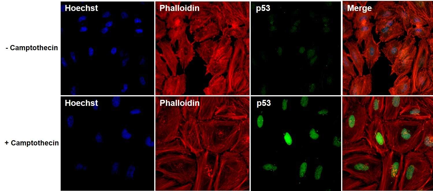
- Immunofluorescent analysis of p53 (green) in untreated and 5uM 20 hours Camptothecin treated HeLa cells. The cells were fixed with 4% paraformaldehyde for 15 minutes at -20c, permeabilized with 0.1% Triton X-100 for 15 minutes, and blocked with 3% BSA for 30 minutes at room temperature. Cells were stained with a p53 mouse monoclonal antibody (Product # MA5-12571) at a concentration of 5 µg/mL in blocking buffer for 1 hour at room temperature, and then incubated with a Goat anti-Mouse IgG (H+L) Secondary Antibody, Alexa Fluor Plus 488 conjugate (Product # A32731) at a dilution of 1:500 for at least 30 minutes at a room temperature in the dark (green). F-actin (red) was stained by Dylight 554 Phalloidin (Product #21834) and nuclei (blue) were stained with Hoechst 33342 (Product # 62249). Images were taken on a Thermo Scientific ToxInsight Instrument at 20X magnification.
Supportive validation
- Submitted by
- Invitrogen Antibodies (provider)
- Main image

- Experimental details
- Immunoprecipitation of p53 using p53 Monoclonal Antibody (MA5-12571) on denatured Human MDA231 Cells.

- Submitted by
- Invitrogen Antibodies (provider)
- Main image

- Experimental details
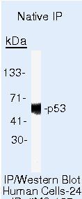
- Immunoprecipitation of p53 using p53 Monoclonal Antibody (MA5-12571) on Native Human SKBR3 Cells.
Supportive validation
- Submitted by
- Invitrogen Antibodies (provider)
- Main image

- Experimental details
- Immunohistochemistry was performed on human ovary, human ovary cancer and human liver. Tissue was deparaffinized with xylene, followed by rehydration in sequential washes of 100% ethanol, 95% ethanol, 80% ethanol, and water. To expose target proteins, antigen retrieval was performed using 10mM sodium citrate (pH 6.0) and heated for 20 min in Lab Vision PT Module (Product # A80400012). Following antigen retrieval, tissues were blocked in a 10% goat serum (Product # 31872) in wash buffer solution for 30 minutes at room temperature and endogenous peroxidase activity quenched with Peroxidase Suppressor (Product # 35000). Tissue was then probed with a p53 mouse monoclonal antibody (Product # MA5-12571) at a concentration of 2.5 µg/mL in 10% goat serum in wash buffer for 1 hour at room temperature in a humidified chamber. Negative control ovarian cancer tissue received no primary antibody. Tissues were washed extensively with PBST, and detection was performed using a SuperBoost™ goat anti-mouse Poly HRP secondary antibody reagent (Product # B40961) followed by colorimetric detection using DAB Quanto (Product # TA-125-QHDX). Tissues were then counterstained with hematoxylin (Product # TA-125-MH), mounted and imaged on EVOS FL Auto Imaging Station.

Supportive validation
- Submitted by
- Invitrogen Antibodies (provider)
- Main image

- Experimental details
- NULL

- Submitted by
- Invitrogen Antibodies (provider)
- Main image

- Experimental details
- NULL

- Submitted by
- Invitrogen Antibodies (provider)
- Main image

- Experimental details
- NULL

- Submitted by
- Invitrogen Antibodies (provider)
- Main image

- Experimental details
- NULL

- Submitted by
- Invitrogen Antibodies (provider)
- Main image

- Experimental details
- NULL

- Submitted by
- Invitrogen Antibodies (provider)
- Main image

- Experimental details
- NULL
- Submitted by
- Invitrogen Antibodies (provider)
- Main image

- Experimental details
- Figure 2 p53 accumulation and activation, caused by HUWE1 p.G4310R, perturb the cell cycle and impair proliferation in JMS patient-derived cells (A) Immunoblot analysis of the HUWE1, p53, and p21 protein levels in healthy control LCs and LCs from two JMS patients (JMS1 and JMS2). p53 protein levels relative to tubulin, serving as loading control, are indicated. (B) In vitro ubiquitination of purified recombinant p53 with increasing amounts of wild-type (WT) and p.G4310R HECT proteins. (C) In silico analysis of difference in the folding free energy change (DeltaDeltaG) of WT and G4310R HUWE1 using the indicated prediction tools. (D) Cell-cycle distribution determined by flow cytometry of healthy control, JMS1, and JMS2 LCs. (E) Fraction of annexin V-positive apoptotic cells measured by flow cytometry. (F) Proliferation rate of healthy control, JMS1, and JMS2 LCs. All error bars indicate mean +- SEM (n >= 3, biological replicates). Statistical significance was determined by two-way ANOVA with Tukey post-test (D) and Bonferroni post-test (F); one-way ANOVA with Bonferroni post-test; *p

- Submitted by
- Invitrogen Antibodies (provider)
- Main image
- Experimental details
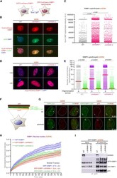
- Figure 5 AHNAK-regulated 53BP1 phase separation culminates in optimal p53 response (A) Schematic illustration of blue light-induced optoDroplet formation by 53BP1. (B) Representative image of Cry2-mCherry-53BP1-MFFR-BRCT-W1495A optoDroplet formation in WT and AHNAK -/- U2OS cells. After optoDroplet induction cells, were fixed and stained for p53 (fluorescein green) and DAPI (blue). (C) Quantification of sum 53BP1 optoDroplet fluorescence intensity in individual cells of the specified genotype. Solid line denotes mean. A.U., arbitrary units (WT, n = 222; DeltaAHNAK-1, n = 1,083; and DeltaAHNAK-2, n = 412). (D) Representative image of Cry2-mCherry optoDroplet formation in WT and AHNAK -/- U2OS cells transfected with CRY2 harboring or not the OD domain. Nuclei are depicted in blue (DAPI). (E) Quantification of sum 53BP1 optoDroplet fluorescence intensity in individual cells of the specified genotype from (D). Solid line denotes median. A.U., arbitrary units (n >= 2,650). (F) Schematic illustration of the LacO-LacI tethering system. (G) Co-localization of control GFP-LacI or GFP-53BP1-LacI (fluorescein green) with p53 (red) on the LacO array in U2OS19 cells after depletion with indicated siRNAs (n = 2 independent experiments). The white numbers represent the percentage of cells that exert colocalization of the indicated protein with the lacO array. (H) Fluorescence recovery after photobleaching (FRAP) analysis on the 53BP1-NBs. Graphical representation for normalized fluorescence

- Submitted by
- Invitrogen Antibodies (provider)
- Main image

- Experimental details
- Figure 6. DNA-RNA hybrid-dependent DSB foci formation and onset of DSB signalling. ( A ) Confocal imaging of MDC1 and GFP-RNaseH1 following transient transfection of pEGFP-M27 or non-transfected (mock) cells. n , number of cells with shown phenotype in %. ( B ) ChIP analysis of HA-53BP1 occupancy at DS1 using HA antibody (3F10) and site-specific primers. Asterisk, P -value < 0.05, two-tailed t -test. Error bar: mean +- SEM, n = 3. ( C ) Immunoblots detecting phospho-ATM, total ATM, gammaH2A.X, phospho-Chk1 and p53 levels following fluorescence-activated cell sorting (FACS) in absence of 4OHT (left) and after pulse-chase 4OHT incubation kinetics (right). Ponceau S, control.
