 Explore
 Validate
 Learn
Learn Western blot
Antibody data
- Antibody Data
- Antigen structure
- References [0]
- Comments [0]
- Validations
- Western blot [2]
- Immunocytochemistry [2]
- Immunohistochemistry [1]
Submit
Validation data
Reference
Comment
Report error
- Product number
- PA5-30947 - Provider product page

- Provider
- Invitrogen Antibodies
- Product name
- Methyl-p53 (Lys372) Polyclonal Antibody
- Antibody type
- Polyclonal
- Antigen
- Synthetic peptide
- Description
- Recommended positive controls: HCT116 24hr cisplatin 30uM, 48hr cisplatin uM. Predicted reactivity: Sheep (100%), Rhesus Monkey (100%). Store product as a concentrated solution. Centrifuge briefly prior to opening the vial.
- Reactivity
- Human
- Host
- Rabbit
- Isotype
- IgG
- Vial size
- 100 µL
- Concentration
- 1.43 mg/mL
- Storage
- Store at 4°C short term. For long term storage, store at -20°C, avoiding freeze/thaw cycles.
No comments: Submit comment
Supportive validation
- Submitted by
- Invitrogen Antibodies (provider)
- Main image

- Experimental details
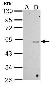
- Western blot analysis of Mono-Methyl-p53 (Lys372) using 30 µg of A) HCT116 cells with mock treatment for 24hr and B) HCT116 cells with 30uM cisplatin treatment for 24hr lysate. Samples were loaded onto a 10% SDS-PAGE gel and probed with a Mono-Methyl-p53 (Lys372) polyclonal antibody (Product # PA5-30947) at a dilution of 1:500.
- Submitted by
- Invitrogen Antibodies (provider)
- Main image

- Experimental details
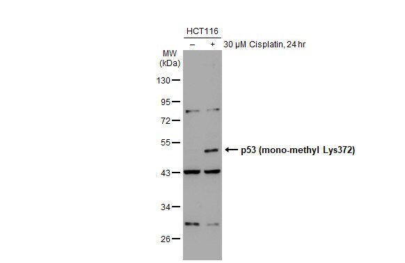
- Western Blot using Methyl-p53 (Lys372) Polyclonal Antibody (Product # PA5-30947). Untreated (–) and treated (+) HCT-116 whole cell extracts (30 µg) were separated by 10% SDS-PAGE, and the membrane was blotted with Methyl-p53 (Lys372) Polyclonal Antibody (Product # PA5-30947) diluted at 1:500. The HRP-conjugated anti-rabbit IgG antibody was used to detect the primary antibody.
Supportive validation
- Submitted by
- Invitrogen Antibodies (provider)
- Main image
- Experimental details
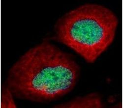
- Immunofluorescent analysis of Mono-Methyl-p53 (Lys372) in paraformaldehyde-fixed U2OS cells using a Mono-Methyl-p53 (Lys372) polyclonal antibody (Product # PA5-30947) (Green) at a 1:500 dilution. Alpha-tubulin filaments were labeled with Product # PA5-29281 (Red) at a 1:2000.

- Submitted by
- Invitrogen Antibodies (provider)
- Main image

- Experimental details
- Immunofluorescence analysis of Acetyl-p53(Lys372) was performed using 70% confluent log phase HeLa cells treated with 0.5uM Doxorubicin for 24 hrs. The cells were fixed with 4% paraformaldehyde for 10 minutes, permeabilized with 0.1% Triton™ X-100 for 10 minutes, and blocked with 1% BSA for 1 hour at room temperature. The cells were labeled with Methyl-p53(Lys 372) Polyclonal Antibody (Product # PA5-30947) at 5µg/mL in 0.1% BSA, incubated overnight at 4 degree Celsius and then labeled with Goat anti-Rabbit IgG (H+L) Superclonal™ Secondary Antibody, Alexa Fluor® 488 conjugate (Product # A27034) at a dilution of 1:2000 for 45 minutes at room temperature (Panel a: green). Nuclei (Panel b: blue) were stained with SlowFade® Gold Antifade Mountant with DAPI (Product # S36938). F-actin (Panel c: red) was stained with Rhodamine Phalloidin (Product # R415, 1:300). Panel d represents the merged image showing nuclear localization. Panel e represents the untreated cells with negligible expression of Methyl-p53 (Lys 372). Panel f shows control cells with no primary antibody to assess background. The images were captured at 60X magnification.

Supportive validation
- Submitted by
- Invitrogen Antibodies (provider)
- Main image
- Experimental details
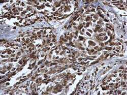
- Immunohistochemistry (Paraffin) analysis of Methyl-p53 (Lys372) was performed in paraffin-embedded human cervical cancer tissue using Methyl-p53 (Lys372) Polyclonal Antibody (Product # PA5-30947) at a dilution of 1:500.
