Explore
 Validate
 Learn
Learn Western blot
 Immunoprecipitation
Antibody data
- Antibody Data
- Antigen structure
- References [11]
- Comments [0]
- Validations
- Western blot [3]
- Immunocytochemistry [1]
- Chromatin Immunoprecipitation [1]
Submit
Validation data
Reference
Comment
Report error
- Product number
- AF1355 - Provider product page

- Provider
- R&D Systems
- Product name
- Human/Mouse/Rat p53 Antibody
- Antibody type
- Polyclonal
- Description
- Antigen Affinity-purified. Detects human, mouse, and rat p53.
- Reactivity
- Human, Mouse, Rat
- Host
- Goat
- Conjugate
- Unconjugated
- Antigen sequence
P04637- Isotype
- IgG
- Vial size
- 100 ug
- Concentration
- LYOPH
- Storage
- Use a manual defrost freezer and avoid repeated freeze-thaw cycles. 12 months from date of receipt, -20 to -70 °C as supplied. 1 month, 2 to 8 °C under sterile conditions after reconstitution. 6 months, -20 to -70 °C under sterile conditions after reconstitution.
Submitted references Progranulin protects the mouse retina under hypoxic conditions via inhibition of the Toll‑like receptor‑4‑NADPH oxidase 4 signaling pathway.
B-cell lymphoma 2 is associated with advanced tumor grade and clinical stage, and reduced overall survival in young Chinese patients with colorectal carcinoma.
Dysfunction of the MDM2/p53 axis is linked to premature aging.
Combining Anti-Mir-155 with Chemotherapy for the Treatment of Lung Cancers.
Chromosomal instability induced by increased BIRC5/Survivin levels affects tumorigenicity of glioma cells.
Growth hormone is permissive for neoplastic colon growth.
Survivin safeguards chromosome numbers and protects from aneuploidy independently from p53.
Expression of S100A6 in cardiac myocytes limits apoptosis induced by tumor necrosis factor-alpha.
Protection of human keratinocytes from UVB-induced inflammation using root extract of Lithospermum erythrorhizon.
Protection of human keratinocytes from UVB-induced inflammation using root extract of Lithospermum erythrorhizon.
TIMP-1 gene deficiency increases tumour cell sensitivity to chemotherapy-induced apoptosis.
You ZP, Yu MJ, Zhang YL, Shi K
Molecular medicine reports 2019 Jan;19(1):382-390
Molecular medicine reports 2019 Jan;19(1):382-390
B-cell lymphoma 2 is associated with advanced tumor grade and clinical stage, and reduced overall survival in young Chinese patients with colorectal carcinoma.
Wang J, He G, Yang Q, Bai L, Jian B, Li Q, Li Z
Oncology letters 2018 Jun;15(6):9009-9016
Oncology letters 2018 Jun;15(6):9009-9016
Dysfunction of the MDM2/p53 axis is linked to premature aging.
Lessel D, Wu D, Trujillo C, Ramezani T, Lessel I, Alwasiyah MK, Saha B, Hisama FM, Rading K, Goebel I, Schütz P, Speit G, Högel J, Thiele H, Nürnberg G, Nürnberg P, Hammerschmidt M, Zhu Y, Tong DR, Katz C, Martin GM, Oshima J, Prives C, Kubisch C
The Journal of clinical investigation 2017 Oct 2;127(10):3598-3608
The Journal of clinical investigation 2017 Oct 2;127(10):3598-3608
Combining Anti-Mir-155 with Chemotherapy for the Treatment of Lung Cancers.
Van Roosbroeck K, Fanini F, Setoyama T, Ivan C, Rodriguez-Aguayo C, Fuentes-Mattei E, Xiao L, Vannini I, Redis RS, D'Abundo L, Zhang X, Nicoloso MS, Rossi S, Gonzalez-Villasana V, Rupaimoole R, Ferracin M, Morabito F, Neri A, Ruvolo PP, Ruvolo VR, Pecot CV, Amadori D, Abruzzo L, Calin S, Wang X, You MJ, Ferrajoli A, Orlowski R, Plunkett W, Lichtenberg TM, Davuluri RV, Berindan-Neagoe I, Negrini M, Wistuba II, Kantarjian HM, Sood AK, Lopez-Berestein G, Keating MJ, Fabbri M, Calin GA
Clinical cancer research : an official journal of the American Association for Cancer Research 2017 Jun 1;23(11):2891-2904
Clinical cancer research : an official journal of the American Association for Cancer Research 2017 Jun 1;23(11):2891-2904
Chromosomal instability induced by increased BIRC5/Survivin levels affects tumorigenicity of glioma cells.
Conde M, Michen S, Wiedemuth R, Klink B, Schröck E, Schackert G, Temme A
BMC cancer 2017 Dec 28;17(1):889
BMC cancer 2017 Dec 28;17(1):889
Growth hormone is permissive for neoplastic colon growth.
Chesnokova V, Zonis S, Zhou C, Recouvreux MV, Ben-Shlomo A, Araki T, Barrett R, Workman M, Wawrowsky K, Ljubimov VA, Uhart M, Melmed S
Proceedings of the National Academy of Sciences of the United States of America 2016 Jun 7;113(23):E3250-9
Proceedings of the National Academy of Sciences of the United States of America 2016 Jun 7;113(23):E3250-9
Survivin safeguards chromosome numbers and protects from aneuploidy independently from p53.
Wiedemuth R, Klink B, Töpfer K, Schröck E, Schackert G, Tatsuka M, Temme A
Molecular cancer 2014 May 9;13:107
Molecular cancer 2014 May 9;13:107
Expression of S100A6 in cardiac myocytes limits apoptosis induced by tumor necrosis factor-alpha.
Tsoporis JN, Izhar S, Parker TG
The Journal of biological chemistry 2008 Oct 31;283(44):30174-83
The Journal of biological chemistry 2008 Oct 31;283(44):30174-83
Protection of human keratinocytes from UVB-induced inflammation using root extract of Lithospermum erythrorhizon.
Ishida T, Sakaguchi I
Biological & pharmaceutical bulletin 2007 May;30(5):928-34
Biological & pharmaceutical bulletin 2007 May;30(5):928-34
Protection of human keratinocytes from UVB-induced inflammation using root extract of Lithospermum erythrorhizon.
Ishida T, Sakaguchi I
Biological & pharmaceutical bulletin 2007 May;30(5):928-34
Biological & pharmaceutical bulletin 2007 May;30(5):928-34
TIMP-1 gene deficiency increases tumour cell sensitivity to chemotherapy-induced apoptosis.
Davidsen ML, Würtz SØ, Rømer MU, Sørensen NM, Johansen SK, Christensen IJ, Larsen JK, Offenberg H, Brünner N, Lademann U
British journal of cancer 2006 Oct 23;95(8):1114-20
British journal of cancer 2006 Oct 23;95(8):1114-20
No comments: Submit comment
Supportive validation
- Submitted by
- R&D Systems (provider)
- Main image
- Experimental details
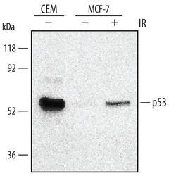
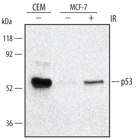
- Detection of Human p53 by Western Blot. Western blot shows lysates of CEM human T-lymphoblastoid cell line and MCF-7 human breast cancer cell line were mock-treated (-) or exposed (+) to 10 Gy ionizing radiation (IR) and harvested after 1 hour. PVDF membrane was probed with 0.5 µg/mL of Goat Anti-Human/Mouse/Rat p53 Antigen Affinity-purified Polyclonal Antibody (Catalog # AF1355), followed by HRP-conjugated Anti-Goat IgG Secondary Antibody (Catalog # HAF109). A specific band was detected for p53 at approximately 53 kDa (as indicated). This experiment was conducted under reducing conditions and using Immunoblot Buffer Group 1.
- Submitted by
- R&D Systems (provider)
- Main image
- Experimental details
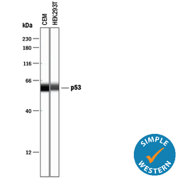
- Detection of Human p53 by Simple WesternTM. Simple Western lane view shows lysates of CEM human T-lymphoblastoid cell line and HEK293T human embryonic kidney cell line, loaded at 0.2 mg/mL. A specific band was detected for p53 at approximately 59 kDa (as indicated) using 2.5 µg/mL of Goat Anti-Human/Mouse/Rat p53 Antigen Affinity-purified Polyclonal Antibody (Catalog # AF1355) followed by 1:50 dilution of HRP-conjugated Anti-Goat IgG Secondary Antibody (Catalog # HAF109). This experiment was conducted under reducing conditions and using the 12-230 kDa separation system.

- Submitted by
- R&D Systems (provider)
- Main image
- Experimental details
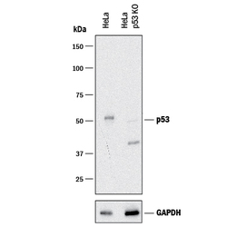
- Western Blot Shows Human p53 Specificity by Using Knockout Cell Line. Western blot shows lysates of HeLa human cervical epithelial carcinoma parental cell line and p53 knockout HeLa cell line (KO). PVDF membrane was probed with 0.25 µg/mL of Goat Anti-Human/Mouse/Rat p53 Antigen Affinity-purified Polyclonal Antibody (Catalog # AF1355) followed by HRP-conjugated Anti-Goat IgG Secondary Antibody (Catalog # HAF017). A specific band was detected for p53 at approximately 53 kDa (as indicated) in the parental HeLa cell line, but is not detectable in knockout HeLa cell line. GAPDH (Catalog # AF5718) is shown as a loading control.This experiment was conducted under reducing conditions and using Immunoblot Buffer Group 1.
Supportive validation
- Submitted by
- R&D Systems (provider)
- Main image

- Experimental details
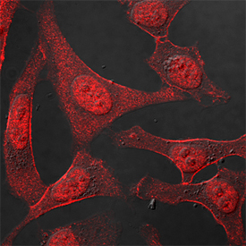
- p53 in HeLa Human Cell Line. p53 was detected in immersion fixed HeLa human cervical epithelial carcinoma cell line using Goat Anti-Human/Mouse/Rat p53 Antigen Affinity-purified Polyclonal Antibody (Catalog # AF1355) at 1.7 µg/mL for 3 hours at room temperature. Cells were stained using the NorthernLights™ 557-conjugated Anti-Goat IgG Secondary Antibody (red; Catalog # NL001) and counterstained with DAPI (blue). Specific staining was localized to nuclei. View our protocol for Fluorescent ICC Staining of Cells on Coverslips.
Supportive validation
- Submitted by
- R&D Systems (provider)
- Main image
- Experimental details
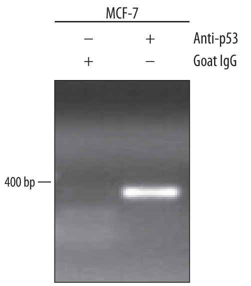
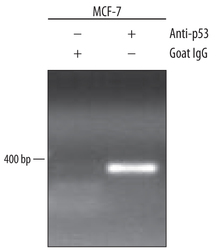
- Detection of p53-regulated Genes by Chromatin Immunoprecipitation. MCF-7 human breast cancer cell line treated with 300 nM camptothecin overnight were fixed using formaldehyde, resuspended in lysis buffer, and sonicated to shear chromatin. p53/DNA complexes were immunoprecipitated using 5 μg Goat Anti-Human/Mouse/Rat p53 Antigen Affinity-purified Polyclonal Antibody (Catalog # AF1355) or control antibody (Catalog # AB-108-C) for 15 minutes in an ultrasonic bath, followed by Biotinylated Anti-Goat IgG Secondary Antibody (Catalog # BAF109). Immunocomplexes were captured using 50 μL of MagCellect Streptavidin Ferrofluid (Catalog # MAG999) and DNA was purified using chelating resin solution. The p21 promoter was detected by standard PCR.