 Explore
 Validate
 Learn
Learn Western blot
 Immunocytochemistry
Antibody data
- Antibody Data
- Antigen structure
- References [1]
- Comments [0]
- Validations
- Immunocytochemistry [1]
Submit
Validation data
Reference
Comment
Report error
- Product number
- HPA027271 - Provider product page

- Provider
- Atlas Antibodies
- Proper citation
- Atlas Antibodies Cat#HPA027271, RRID:AB_10602004
- Product name
- Anti-WDR77
- Antibody type
- Polyclonal
- Description
- Polyclonal Antibody against Human WDR77, Gene description: WD repeat domain 77, Alternative Gene Names: MEP50, Validated applications: ICC, IHC, WB, Uniprot ID: Q9BQA1, Storage: Store at +4°C for short term storage. Long time storage is recommended at -20°C.
- Reactivity
- Human, Mouse, Rat
- Host
- Rabbit
- Conjugate
- Unconjugated
- Isotype
- IgG
- Vial size
- 100 µl
- Concentration
- 0.2 mg/ml
- Storage
- Store at +4°C for short term storage. Long time storage is recommended at -20°C.
- Handling
- The antibody solution should be gently mixed before use.
Submitted references Proteomics and C9orf72 neuropathology identify ribosomes as poly-GR/PR interactors driving toxicity.
Hartmann H, Hornburg D, Czuppa M, Bader J, Michaelsen M, Farny D, Arzberger T, Mann M, Meissner F, Edbauer D
Life science alliance 2018 May;1(2):e201800070
Life science alliance 2018 May;1(2):e201800070
No comments: Submit comment
Supportive validation
- Submitted by
- Atlas Antibodies (provider)
- Main image
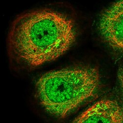
- Experimental details
- Immunofluorescent staining of human cell line A-431 shows localization to nucleoplasm, cytosol & the Golgi apparatus.
- Sample type
- Human
