Explore
 Validate
 Learn
Learn Western blot
 ELISA
ELISAAntibody data
- Antibody Data
- Antigen structure
- References [15]
- Comments [0]
- Validations
- Western blot [1]
- Immunocytochemistry [1]
Submit
Validation data
Reference
Comment
Report error
- Product number
- 15361-1-AP - Provider product page

- Provider
- Proteintech Group
- Proper citation
- Proteintech Cat#15361-1-AP, RRID:AB_2060227
- Product name
- Beta Arrestin 1 antibody
- Antibody type
- Polyclonal
- Description
- KD/KO validated Beta Arrestin 1 antibody (Cat. #15361-1-AP) is a rabbit polyclonal antibody that shows reactivity with human, mouse, rat and has been validated for the following applications: IHC, IP, WB, ELISA.
- Reactivity
- Human, Mouse, Rat
- Host
- Rabbit
- Conjugate
- Unconjugated
- Isotype
- IgG
- Vial size
- 20ul, 150ul
Submitted references Insufficient BK channel function enhances NF-κB nuclear translocation and promotes IL-6 synthesis in vascular smooth muscle cells induced by AT1-AA.
NPAS2, transcriptionally activated by ARRB1, promotes the malignant behaviours of lung adenocarcinoma cells and regulates the reprogramming of glucose metabolism.
Blue light irradiation inhibits the M2 polarization of the cancer-associated macrophages in colon cancer.
Identification and validation of chemokine system-related genes in idiopathic pulmonary fibrosis.
NE-activated β(2)-AR/β-arrestin 2/Src pathway mediates duodenal hyperpermeability induced by water-immersion restraint stress.
Hepatocyte-derived MASP1-enriched small extracellular vesicles activate HSCs to promote liver fibrosis.
CXCL12/CXCR7/β-arrestin1 biased signal promotes epithelial-to-mesenchymal transition of colorectal cancer by repressing miRNAs through YAP1 nuclear translocation.
PLEKHH2 binds β-arrestin1 through its FERM domain, activates FAK/PI3K/AKT phosphorylation, and promotes the malignant phenotype of non-small cell lung cancer.
Blue light irradiation inhibits the growth of colon cancer and activation of cancer‑associated fibroblasts.
Autonomous sensing of the insulin peptide by an olfactory G protein-coupled receptor modulates glucose metabolism.
An ionic lock and a hydrophobic zipper mediate the coupling between an insect pheromone receptor BmOR3 and downstream effectors.
Bioactive peptide apelin rescues acute kidney injury by protecting the function of renal tubular mitochondria.
BBSome trains remove activated GPCRs from cilia by enabling passage through the transition zone.
β-Arrestins promote podocyte injury by inhibition of autophagy in diabetic nephropathy.
Cell survival following radiation exposure requires miR-525-3p mediated suppression of ARRB1 and TXN1.
Li Y, Xue L, Feng J, Wang Z, Long Y, Liu W, Zhang S, Zhi X, Hao H, Wang X, Liu H, Wang L
Biochemical pharmacology 2025 Sep;239:117000
Biochemical pharmacology 2025 Sep;239:117000
NPAS2, transcriptionally activated by ARRB1, promotes the malignant behaviours of lung adenocarcinoma cells and regulates the reprogramming of glucose metabolism.
Wang S, Huang C, Zheng Y, Wu X, Zhong Y
Clinical and experimental pharmacology & physiology 2024 May;51(5):e13860
Clinical and experimental pharmacology & physiology 2024 May;51(5):e13860
Blue light irradiation inhibits the M2 polarization of the cancer-associated macrophages in colon cancer.
Yoshimoto T, Nishi M, Okikawa S, Yoshikawa K, Tokunaga T, Nakao T, Takasu C, Kashihara H, Wada Y, Noma T, Shimada M
BMC cancer 2024 May 31;24(1):664
BMC cancer 2024 May 31;24(1):664
Identification and validation of chemokine system-related genes in idiopathic pulmonary fibrosis.
Zhao T, Wu X, Zhao X, Yao K, Li X, Ni J
Frontiers in immunology 2023;14:1159856
Frontiers in immunology 2023;14:1159856
NE-activated β(2)-AR/β-arrestin 2/Src pathway mediates duodenal hyperpermeability induced by water-immersion restraint stress.
Yan JT, Zhu YZ, Liang L, Feng XY
American journal of physiology. Cell physiology 2023 Jan 1;324(1):C133-C141
American journal of physiology. Cell physiology 2023 Jan 1;324(1):C133-C141
Hepatocyte-derived MASP1-enriched small extracellular vesicles activate HSCs to promote liver fibrosis.
Liu X, Tan S, Liu H, Jiang J, Wang X, Li L, Wu B
Hepatology (Baltimore, Md.) 2023 Apr 1;77(4):1181-1197
Hepatology (Baltimore, Md.) 2023 Apr 1;77(4):1181-1197
CXCL12/CXCR7/β-arrestin1 biased signal promotes epithelial-to-mesenchymal transition of colorectal cancer by repressing miRNAs through YAP1 nuclear translocation.
Si M, Song Y, Wang X, Wang D, Liu X, Qu X, Song Z, Yu X
Cell & bioscience 2022 Oct 9;12(1):171
Cell & bioscience 2022 Oct 9;12(1):171
PLEKHH2 binds β-arrestin1 through its FERM domain, activates FAK/PI3K/AKT phosphorylation, and promotes the malignant phenotype of non-small cell lung cancer.
Wang R, Wang S, Li Z, Luo Y, Zhao Y, Han Q, Rong XZ, Guo YX, Liu Y
Cell death & disease 2022 Oct 8;13(10):858
Cell death & disease 2022 Oct 8;13(10):858
Blue light irradiation inhibits the growth of colon cancer and activation of cancer‑associated fibroblasts.
Yoshimoto T, Shimada M, Tokunaga T, Nakao T, Nishi M, Takasu C, Kashihara H, Wada Y, Okikawa S, Yoshikawa K
Oncology reports 2022 May;47(5)
Oncology reports 2022 May;47(5)
Autonomous sensing of the insulin peptide by an olfactory G protein-coupled receptor modulates glucose metabolism.
Cheng J, Yang Z, Ge XY, Gao MX, Meng R, Xu X, Zhang YQ, Li RZ, Lin JY, Tian ZM, Wang J, Ning SL, Xu YF, Yang F, Gu JK, Sun JP, Yu X
Cell metabolism 2022 Feb 1;34(2):240-255.e10
Cell metabolism 2022 Feb 1;34(2):240-255.e10
An ionic lock and a hydrophobic zipper mediate the coupling between an insect pheromone receptor BmOR3 and downstream effectors.
Lin JY, Yang Z, Yang C, Du JX, Yang F, Cheng J, Pan W, Zhang SJ, Yan X, Wang J, Wang J, Tie L, Yu X, Chen X, Sun JP
The Journal of biological chemistry 2021 Oct;297(4):101160
The Journal of biological chemistry 2021 Oct;297(4):101160
Bioactive peptide apelin rescues acute kidney injury by protecting the function of renal tubular mitochondria.
Guan YM, Diao ZL, Huang HD, Zheng JF, Zhang QD, Wang LY, Liu WH
Amino acids 2021 Aug;53(8):1229-1240
Amino acids 2021 Aug;53(8):1229-1240
BBSome trains remove activated GPCRs from cilia by enabling passage through the transition zone.
Ye F, Nager AR, Nachury MV
The Journal of cell biology 2018 May 7;217(5):1847-1868
The Journal of cell biology 2018 May 7;217(5):1847-1868
β-Arrestins promote podocyte injury by inhibition of autophagy in diabetic nephropathy.
Liu J, Li QX, Wang XJ, Zhang C, Duan YQ, Wang ZY, Zhang Y, Yu X, Li NJ, Sun JP, Yi F
Cell death & disease 2016 Apr 7;7(4):e2183
Cell death & disease 2016 Apr 7;7(4):e2183
Cell survival following radiation exposure requires miR-525-3p mediated suppression of ARRB1 and TXN1.
Kraemer A, Barjaktarovic Z, Sarioglu H, Winkler K, Eckardt-Schupp F, Tapio S, Atkinson MJ, Moertl S
PloS one 2013;8(10):e77484
PloS one 2013;8(10):e77484
No comments: Submit comment
Supportive validation
- Submitted by
- Proteintech Group (provider)
- Main image
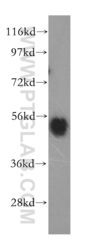
- Experimental details
- mouse lung tissue were subjected to SDS PAGE followed by western blot with 15361-1-AP(ARRB1 antibody) at dilution of 1:500
- Sample type
- tissue

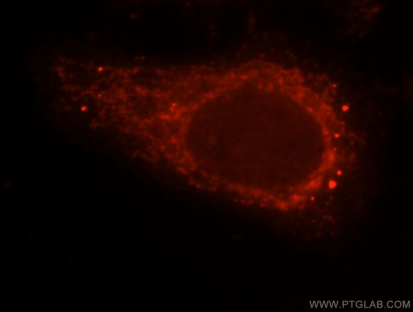
Supportive validation
- Submitted by
- Proteintech Group (provider)
- Main image

- Experimental details
- Immunofluorescent analysis of HepG2 cells, using ARRB1 antibody 15361-1-AP at 1:25 dilution and Rhodamine-labeled goat anti-rabbit IgG (red).
- Sample type
- cell line