 Explore
 Validate
 Learn
Learn Immunocytochemistry
Antibody data
- Antibody Data
- Antigen structure
- References [0]
- Comments [0]
- Validations
- Immunocytochemistry [1]
Submit
Validation data
Reference
Comment
Report error
- Product number
- 702231 - Provider product page

- Provider
- Invitrogen Antibodies
- Product name
- Anti-CCR3 Antibody (23H17L1), ABfinity™ Rabbit Monoclonal
- Antibody type
- Monoclonal
- Antigen
- Synthetic peptide
- Description
- ABfinity recombinant antibodies are rabbit monoclonal antibodies, unmatched for producing superior results. ABfinity antibodies are developed by immunizing animals, screening for functionality, cloning the immunogen-specific antibody genes into high-level mammalian expression vectors, produced on a large scale, and purified with Protein A. ABfinity monoclonal antibodies resemble rabbit monoclonals isolated from serum or produced by hybridomas, but demonstrate greater specificity and sensitivity. Because ABfinity recombinant antibodies are derived from cloned DNA sequences of the heavy and light antibody chains, they are not susceptible to cell-line drift or lot-to-lot variation, thus allowing for peak specificity and performance. This antibody is predicted to react with Monkey
- Reactivity
- Human
- Host
- Rabbit
- Isotype
- IgG
- Antibody clone number
- 23H17L1
- Vial size
- 100 µg
- Concentration
- 0.5 mg/ml
- Storage
- -20° C, Avoid Freeze/Thaw Cycles
No comments: Submit comment
Supportive validation
- Submitted by
- Invitrogen Antibodies (provider)
- Main image
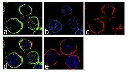
- Experimental details
- For immunofluorescence analysis, Raji cells were fixed and permeabilized for detection of endogenous CCR3 using ABfinity Anti-CCR3 Recombinant Rabbit Monoclonal Antibody (Product # 702231, 2 µg/ml) and labeled with Goat anti-Rabbit IgG (H+L) Superclonal Secondary Antibody, Alexa Fluor® 488 conjugate (Product # A27034, 1:2000). Panel a) shows representative cells that were stained for detection and localization of CCR3 protein (green), Panel b) is stained for nuclei (blue) using SlowFade® Gold Antifade Mountant with DAPI (Product # S36938). Panel c) represents cytoskeletal F-actin staining using Rhodamine Phalloidin (Product # R415, 1:300). Panel d) is a composite image of Panels a, b and c clearly demonstrating membrane localization of CCR3. Panel e) represents control cells with no primary antibody to assess background. The images were captured at 60X magnification.
