 Explore
 Validate
 Learn
LearnMA5-15172
antibody from Invitrogen Antibodies
Targeting: JUN
AP-1, c-Jun
 Western blot Immunocytochemistry
 Immunoprecipitation Immunohistochemistry Chromatin Immunoprecipitation Other assay
Antibody data
- Antibody Data
- Antigen structure
- References [1]
- Comments [0]
- Validations
- Western blot [2]
- Immunocytochemistry [2]
- Immunohistochemistry [1]
- Other assay [1]
Submit
Validation data
Reference
Comment
Report error
- Product number
- MA5-15172 - Provider product page

- Provider
- Invitrogen Antibodies
- Product name
- c-Jun Monoclonal Antibody (C.238.2)
- Antibody type
- Monoclonal
- Antigen
- Other
- Description
- It is not recommended to aliquot this antibody.
- Reactivity
- Human, Mouse, Rat
- Host
- Rabbit
- Isotype
- IgG
- Antibody clone number
- C.238.2
- Vial size
- 100 μL
- Concentration
- 48 μg/mL
- Storage
- -20°C
Submitted references Promoter G-quadruplexes and transcription factors cooperate to shape the cell type-specific transcriptome.
Lago S, Nadai M, Cernilogar FM, Kazerani M, Domíniguez Moreno H, Schotta G, Richter SN
Nature communications 2021 Jun 23;12(1):3885
Nature communications 2021 Jun 23;12(1):3885
No comments: Submit comment
Supportive validation
- Submitted by
- Invitrogen Antibodies (provider)
- Main image
- Experimental details
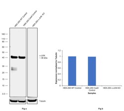
- Knockout of c-JUN was achieved by CRISPR-Cas9 genome editing using LentiArray™ Lentiviral sgRNA (Product # A32042, Assay ID CRISPR905187_SGM) and LentiArray Cas9 Lentivirus (Product # A32064). Western blot analysis of c-JUN was performed by loading 30 µg of HEK-293 wild type (Lane 1), HEK-293 Cas9 (Lane 2) and HEK-293 c-JUN KO (Lane 3) modified whole cell extracts. The samples were electrophoresed using NuPAGE™ Novex™ 4-12% Bis-Tris Protein Gel (Product # NP0321BOX). Resolved proteins were then transferred onto a nitrocellulose membrane (Product # IB23001) by iBlot® 2 Dry Blotting System (Product # IB21001). The blot was probed with c-Jun Monoclonal Antibody (C.238.2) (Product # MA5-15172, 1:1000 dilution) and Goat anti-Rabbit IgG (Heavy Chain) Superclonal™ Recombinant Secondary Antibody, HRP (Product # A27036, 1:10,000 dilution) using the iiBright™ FL1500 (Product # A44115). Chemiluminescent detection was performed using SuperSignal™ West Dura Extended Duration Substrate (Product # 34076). Loss of signal upon CRISPR mediated knockout (KO) using the LentiArray™ CRISPR product line confirms that antibody is specific to c-JUN. An uncharacterized band was observed in HEK-293 WT sample t ~25 kDa.
- Submitted by
- Invitrogen Antibodies (provider)
- Main image

- Experimental details
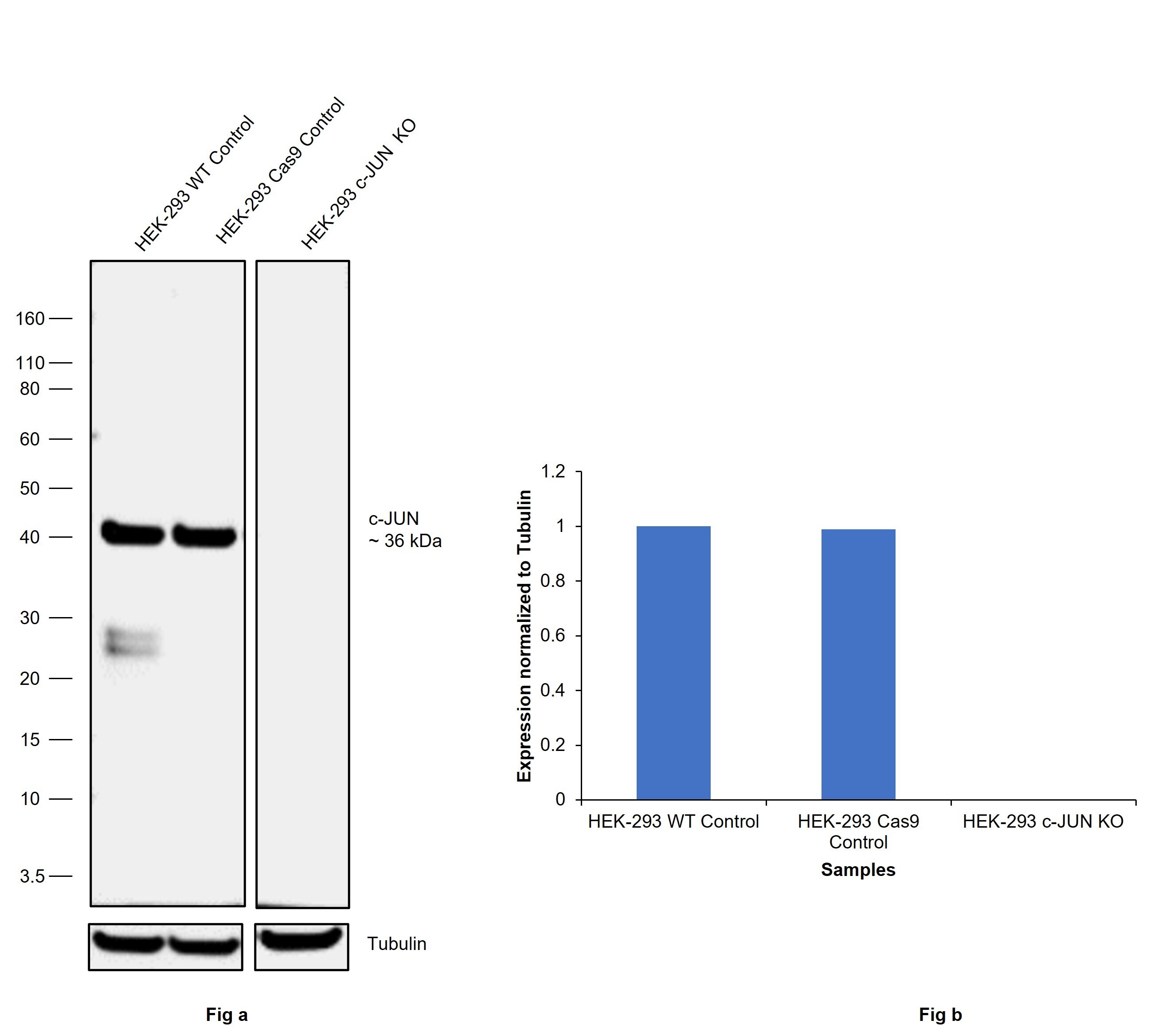
- Western blot analysis was performed on nuclear enriched cell extracts (30 µg lysate) of HeLa (Lane 1), HeLa treated with PMA (50 ng/mL for 2 hours) (Lane 2), HeLa treated with PMA (50 ng/mL for 2 hours) and Trichostatin A (1uM for 1 hour) (Lane 3), U-87 MG (Lane 4) and PC-3 (Lane 5). The blot was probed with Anti-c-Jun Monoclonal Antibody (C.238.2) (Product # MA5-15172, 1:500 dilution) and detected by chemiluminescence using Goat anti-Rabbit IgG (Heavy Chain) Superclonal™ Secondary Antibody, HRP conjugate (Product # A27036, 0.25 µg/mL, 1:4000 dilution). A 40 kDa band corresponding to c-Fos was observed across the cell lines tested and was enhanced upon treatment.
Supportive validation
- Submitted by
- Invitrogen Antibodies (provider)
- Main image

- Experimental details
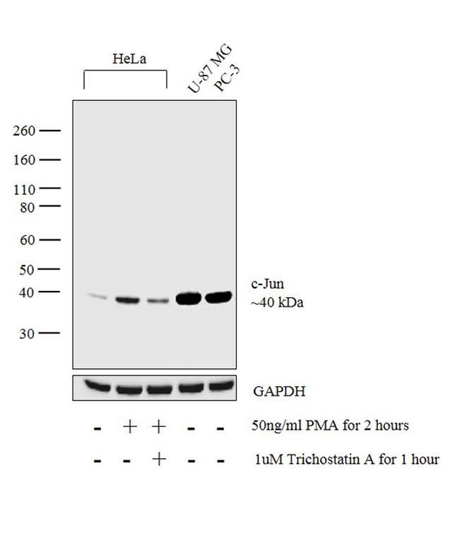
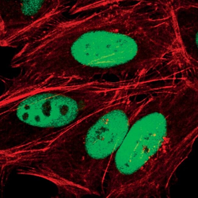
- Immunofluorescent analysis of c-Jun in HeLa cells using a c-Jun monoclonal antibody (Product # MA5-15172) (green). Actin filaments are labeled with a fluorescent red phalloidin.

- Submitted by
- Invitrogen Antibodies (provider)
- Main image
- Experimental details
- Immunofluorescent analysis of c-Jun in HeLa cells using a c-Jun monoclonal antibody (Product # MA5-15172) (green). Actin filaments are labeled with a fluorescent red phalloidin.
Supportive validation
- Submitted by
- Invitrogen Antibodies (provider)
- Main image
- Experimental details
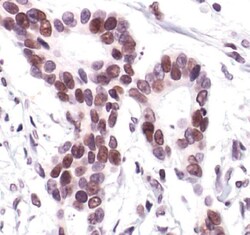
- Immunohistochemical analysis of c-Jun in paraffin-embedded human colon carcinoma using a c-Jun monoclonal antibody (Product # MA5-15172).

Supportive validation
- Submitted by
- Invitrogen Antibodies (provider)
- Main image
- Experimental details
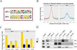
- Fig. 5 Identification of TFs binding to BG4-IP regions in 93T449 cells. A Consensus sequences of TFBSs that are significantly enriched in BG4 ChIP peaks, as calculated by Homer software. AP-1: p value 1e-490, SP1: p value 1e-172. B Position and frequency of TFBSs with respect to the BG4 ChIP peak center. Data points for each TFBS occurrence are reported and fitted according to a nonparametric spline regression curve, the gray area surrounding the curve represents the confidence interval as measure of the regression likelihood. C Percentage of genes with (yellow) and without (darkblue) ChIP-G4s, oG4s, and pG4s containing validated TFBS for AP-1 and SP1, according to ENCODE database. D Western blot showing co-immunoprecipitation of G4s and the two TFs SP1 and AP-1. The INPUT lane 1 corresponds to the total fraction of the sheared chromatin used as starting material. G4s were immunoprecipitated by BG4 antibody (IP BG4), and AP-1 and SP1 were detected by immunoblotting (lane 2). AP-1 (lanes 4 and 5) and SP1 (lanes 6 and 7) TFs were immunoprecipitated from the sheared chromatin with or without previous incubation in the presence of BG4 antibody. Mock G (lane 3) and A (lane 8) are the negative controls immunoprecipitated in the absence of primary antibody using protein-G- or protein-A-coated beads, respectively. The shown blots belong to one of at least two independent biological replicates performed for each sample. Source data for each panel are provided or referenced in the Sour
