Explore
 Validate
 Learn
Learn Western blot
 ELISA
ELISA Immunocytochemistry
Antibody data
- Antibody Data
- Antigen structure
- References [3]
- Comments [0]
- Validations
- Immunocytochemistry [2]
- Immunohistochemistry [2]
- Flow cytometry [3]
- Other assay [2]
Submit
Validation data
Reference
Comment
Report error
- Product number
- MA5-15881 - Provider product page

- Provider
- Invitrogen Antibodies
- Product name
- c-Jun Monoclonal Antibody (5B1)
- Antibody type
- Monoclonal
- Antigen
- Purifed from natural sources
- Description
- MA5-15881 targets c-Jun in indirect ELISA, FACS, IF, IHC, and WB applications and shows reactivity with Human, mouse, and Non-human primate samples. The MA5-15881 immunogen is purified recombinant fragment of human c-Jun expressed in E. Coli. . MA5-15881 detects c-Jun which has a predicted molecular weight of approximately 43kDa.
- Reactivity
- Human, Mouse
- Host
- Mouse
- Isotype
- IgG
- Antibody clone number
- 5B1
- Vial size
- 100 μL
- Concentration
- Conc. not determined
- Storage
- Store at 4°C short term. For long term storage, store at -20°C, avoiding freeze/thaw cycles.
Submitted references Mechanical Study of Jian-Gan-Xiao-Zhi Decoction on Nonalcoholic Fatty Liver Disease Based on Integrated Network Pharmacology and Untargeted Metabolomics.
Estradiol/GPER affects the integrity of mammary duct-like structures in vitro.
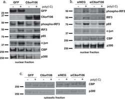
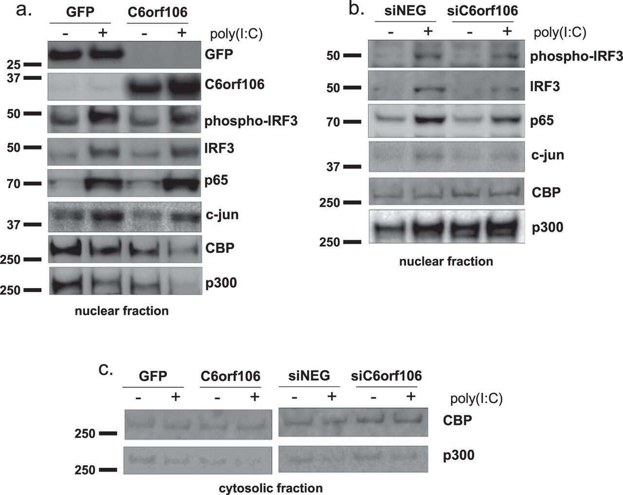
C6orf106 is a novel inhibitor of the interferon-regulatory factor 3-dependent innate antiviral response.
Cao YJ, Li HZ, Zhao J, Sun YM, Jin XW, Lv SQ, Luo JY, Fang XX, Wen WB, Liao JB
Evidence-based complementary and alternative medicine : eCAM 2022;2022:2264394
Evidence-based complementary and alternative medicine : eCAM 2022;2022:2264394
Estradiol/GPER affects the integrity of mammary duct-like structures in vitro.
Deng Y, Miki Y, Nakanishi A
Scientific reports 2020 Jan 28;10(1):1386
Scientific reports 2020 Jan 28;10(1):1386
C6orf106 is a novel inhibitor of the interferon-regulatory factor 3-dependent innate antiviral response.
Ambrose RL, Liu YC, Adams TE, Bean AGD, Stewart CR
The Journal of biological chemistry 2018 Jul 6;293(27):10561-10573
The Journal of biological chemistry 2018 Jul 6;293(27):10561-10573
No comments: Submit comment
Supportive validation
- Submitted by
- Invitrogen Antibodies (provider)
- Main image
- Experimental details
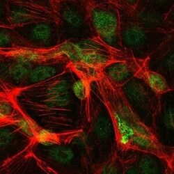
- Immunofluorescence analysis of PC-2 cells using c-Jun monoclonal antibody (Product # MA5-15881) (Green). Red: actin filaments have been labeled with phalloidin.
- Submitted by
- Invitrogen Antibodies (provider)
- Main image

- Experimental details
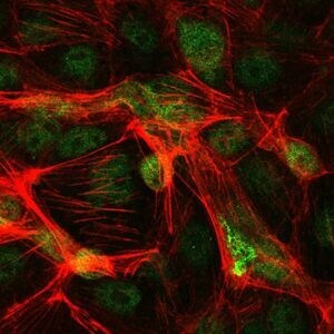
- Immunofluorescence analysis of PC-2 cells using c-Jun monoclonal antibody (Product # MA5-15881) (Green). Red: actin filaments have been labeled with phalloidin.

Supportive validation
- Submitted by
- Invitrogen Antibodies (provider)
- Main image
- Experimental details
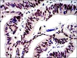
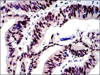
- Immunohistochemical analysis of paraffin-embedded human rectum cancer tissues using c-Jun monoclonal antibody (Product # MA5-15881) followed with DAB staining.
- Submitted by
- Invitrogen Antibodies (provider)
- Main image

- Experimental details
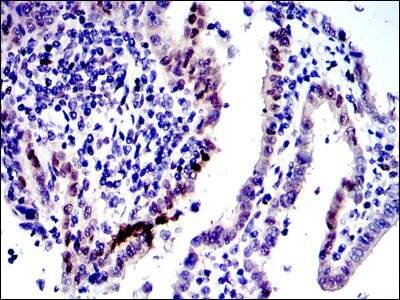
- Immunohistochemical analysis of paraffin-embedded human Tunica intima cancer tissues using c-Jun monoclonal antibody (Product # MA5-15881) followed with DAB staining.
Supportive validation
- Submitted by
- Invitrogen Antibodies (provider)
- Main image

- Experimental details
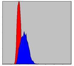
- Flow cytometric analysis of HepG2 cells using c-Jun monoclonal antibody (Product # MA5-15881) (blue) and negative control (red).
- Submitted by
- Invitrogen Antibodies (provider)
- Main image

- Experimental details
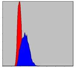
- Flow cytometric analysis of HepG2 cells using c-Jun monoclonal antibody (Product # MA5-15881) (blue) and negative control (red).

- Submitted by
- Invitrogen Antibodies (provider)
- Main image

- Experimental details
- Flow cytometric analysis of HepG2 cells using c-Jun monoclonal antibody (Product # MA5-15881) (blue) and negative control (red).
Supportive validation
- Submitted by
- Invitrogen Antibodies (provider)
- Main image
- Experimental details
- NULL
- Submitted by
- Invitrogen Antibodies (provider)
- Main image

- Experimental details
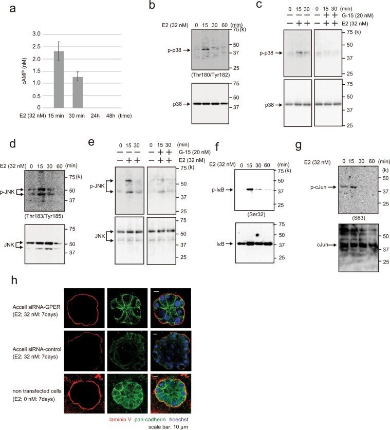
- Figure 2 Analysis of E2 signal transduction. ( a ) cAMP assay showing cAMP levels (nM) in MCF-10A cells following treatment with 32 nM E2 for 15 min, 30 min, 24 h, and 48 h. Three independent experiments were performed. Bars represent +/-SD. ( b ) Western blotting of MCF-10A cells showing p38 and phospho-p38 (Thr180/Tyr182) following treatment with 32 nM E2 for 0-60 min. ( c ) Western blotting of MCF-10A cells treated with 32 nM E2 (left panel) or with 32 nM E2 and 20 nM G-15 (right panel) for 0-30 min. ( d ) Western blotting of MCF-10A cells showing JNK and phosphor-JNK (Thr183/Tyr185) following treatment with 32 nM E2 for 0-60 min. ( e ) Western blotting of MCF-10A cells treated with 32 nM E2 (left panel) or with 32 nM E2 and 20 nM G-15 (right panel) for 0-30 min. ( f ) Western blotting of MCF-10A cells treated with 32 nM E2 for 0-60 min showing IkB and phospho-IkB (Ser32, Ser36). ( g ) Western blotting of MCF-10A cells treated with 32 nM E2 for 0-60 min showing c-Jun and phospho-c-Jun (Ser63). ( h ) Representative confocal images of Accell siRNA-GPER- or siRNA-control-transfected MCF-10A cells in a 3D culture through the middle acini, which were treated with E2 (32 nM, left panels) or control (0 nM, right panel) for 7 days. Laminin V (red); pan-cadherin (green). Scale bars = 20 mum. The presented blots were cropped. Full-length blots are presented in Supplementary Fig. 5 .