Explore
 Validate
 Learn
Learn Western blot
 ELISA
ELISA Immunocytochemistry
Antibody data
- Antibody Data
- Antigen structure
- References [5]
- Comments [0]
- Validations
- Immunocytochemistry [6]
- Immunoprecipitation [1]
- Immunohistochemistry [2]
- Chromatin Immunoprecipitation [2]
- Other assay [3]
Submit
Validation data
Reference
Comment
Report error
- Product number
- MA1-083 - Provider product page

- Provider
- Invitrogen Antibodies
- Product name
- CREB Monoclonal Antibody (LB9)
- Antibody type
- Monoclonal
- Antigen
- Recombinant full-length protein
- Description
- MA1-083 detects the CREB in human, monkey, rat and mouse cells. MA1-083 has been successfully used in immunofluorescence, Western blot, immunoprecipitation, immunohistochemistry, and ELISA procedures. By Western blot, this antibody detects a ~43 kDa protein. The MA1-083 immunogen is human recombinant CREB-A protein.
- Reactivity
- Human, Mouse, Rat
- Host
- Mouse
- Isotype
- IgG
- Antibody clone number
- LB9
- Vial size
- 100 μg
- Concentration
- 1 mg/mL
- Storage
- -20°C, Avoid Freeze/Thaw Cycles
Submitted references Lamin B1 Accumulation's Effects on Autosomal Dominant Leukodystrophy (ADLD): Induction of Reactivity in the Astrocytes.
The transcription factor CREB acts as an important regulator mediating oxidative stress-induced apoptosis by suppressing αB-crystallin expression.
Chenodeoxycholic Acid Ameliorates AlCl(3)-Induced Alzheimer's Disease Neurotoxicity and Cognitive Deterioration via Enhanced Insulin Signaling in Rats.
S-nitrosylation of E3 ubiquitin-protein ligase RNF213 alters non-canonical Wnt/Ca+2 signaling in the P301S mouse model of tauopathy.
Mitochondrial remodeling in mice with cardiomyocyte-specific lipid overload.
Ratti S, Rusciano I, Mongiorgi S, Neri I, Cappellini A, Cortelli P, Suh PG, McCubrey JA, Manzoli L, Cocco L, Ramazzotti G
Cells 2021 Sep 28;10(10)
Cells 2021 Sep 28;10(10)
The transcription factor CREB acts as an important regulator mediating oxidative stress-induced apoptosis by suppressing αB-crystallin expression.
Wang L, Nie Q, Gao M, Yang L, Xiang JW, Xiao Y, Liu FY, Gong XD, Fu JL, Wang Y, Nguyen QD, Liu Y, Liu M, Li DW
Aging 2020 Jun 17;12(13):13594-13617
Aging 2020 Jun 17;12(13):13594-13617
Chenodeoxycholic Acid Ameliorates AlCl(3)-Induced Alzheimer's Disease Neurotoxicity and Cognitive Deterioration via Enhanced Insulin Signaling in Rats.
Bazzari FH, Abdallah DM, El-Abhar HS
Molecules (Basel, Switzerland) 2019 May 24;24(10)
Molecules (Basel, Switzerland) 2019 May 24;24(10)
S-nitrosylation of E3 ubiquitin-protein ligase RNF213 alters non-canonical Wnt/Ca+2 signaling in the P301S mouse model of tauopathy.
Amal H, Gong G, Gjoneska E, Lewis SM, Wishnok JS, Tsai LH, Tannenbaum SR
Translational psychiatry 2019 Jan 29;9(1):44
Translational psychiatry 2019 Jan 29;9(1):44
Mitochondrial remodeling in mice with cardiomyocyte-specific lipid overload.
Elezaby A, Sverdlov AL, Tu VH, Soni K, Luptak I, Qin F, Liesa M, Shirihai OS, Rimer J, Schaffer JE, Colucci WS, Miller EJ
Journal of molecular and cellular cardiology 2015 Feb;79:275-83
Journal of molecular and cellular cardiology 2015 Feb;79:275-83
No comments: Submit comment
Supportive validation
- Submitted by
- Invitrogen Antibodies (provider)
- Main image
- Experimental details
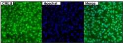
- Immunofluorescent analysis of CREB (green) in untreated HeLa cells. Formalin fixed cells were permeabilized with 0.1% Triton X-100 in TBS for 15 minutes at room temperature. Cells were then blocked with 5% normal goat serum (Product # 31873) for 15 minutes at room temperature. Cells were probed with a mouse monoclonal antibody recognizing CREB (Product # MA1-083), at a dilution of 1:500 for at least 1 hour at room temperature. Cells were washed with PBS and incubated with DyLight 488 goat-anti-mouse secondary antibody at a dilution of 1:400 for 30 minutes at room temperature. Nuclei were stained with Hoechst 33342 dye (Product # 62249). Images were taken on a Thermo Scientific ArrayScan at 20X magnification.

- Submitted by
- Invitrogen Antibodies (provider)
- Main image
- Experimental details
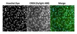
- Immunofluorescent analysis of CREB (green) in untreated HeLa cells. Formalin fixed cells were permeabilized with 0.1% Triton X-100 in TBS for 15 minutes at room temperature. Cells were then blocked with 5% normal goat serum (Product # 31873) for 15 minutes at room temperature. Cells were probed with a mouse monoclonal antibody recognizing CREB (Product # MA1-083), at a dilution of 1:400 for at least 1 hour at room temperature. Cells were washed with PBS and incubated with DyLight 488 goat-anti-mouse secondary antibody at a dilution of 1:400 for 30 minutes at room temperature. Nuclei were stained with Hoechst 33342 dye (Product # 62249). Images were taken on a Thermo Scientific ArrayScan at 20X magnification.

- Submitted by
- Invitrogen Antibodies (provider)
- Main image
- Experimental details
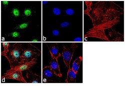
- Immunofluorescence analysis of CREB was performed using 70% confluent log phase MDA-MB-231 cells. The cells were fixed with 4% paraformaldehyde for 10 minutes, permeabilized with 0.1% Triton™ X-100 for 10 minutes, and blocked with 1% BSA for 1 hour at room temperature. The cells were labeled with CREB Monoclonal Antibody (Product # MA1-083) at 5µg/mL in 0.1% BSA and incubated for 3 hours at room temperature and then labeled with Goat anti-Mouse IgG (H+L) Superclonal™ Secondary Antibody, Alexa Fluor® 488 conjugate (Product # A28175) at a dilution of 1:2000 for 45 minutes at room temperature (Panel a: green). Nuclei (Panel b: blue) were stained with SlowFade® Gold Antifade Mountant with DAPI (Product # S36938). F-actin (Panel c: red) was stained with Rhodamine Phalloidin (Product # R415, 1:300). Panel d represents the merged image showing nuclear localization. Panel e shows the no primary antibody control. The images were captured at 60X magnification.

- Submitted by
- Invitrogen Antibodies (provider)
- Main image

- Experimental details
- Immunofluorescent analysis of CREB (green) in untreated HeLa cells. Formalin fixed cells were permeabilized with 0.1% Triton X-100 in TBS for 15 minutes at room temperature. Cells were then blocked with 5% normal goat serum (Product # 31873) for 15 minutes at room temperature. Cells were probed with a mouse monoclonal antibody recognizing CREB (Product # MA1-083), at a dilution of 1:400 for at least 1 hour at room temperature. Cells were washed with PBS and incubated with DyLight 488 goat-anti-mouse secondary antibody at a dilution of 1:400 for 30 minutes at room temperature. Nuclei were stained with Hoechst 33342 dye (Product # 62249). Images were taken on a Thermo Scientific ArrayScan at 20X magnification.

- Submitted by
- Invitrogen Antibodies (provider)
- Main image

- Experimental details
- Immunofluorescent analysis of CREB (green) in untreated HeLa cells. Formalin fixed cells were permeabilized with 0.1% Triton X-100 in TBS for 15 minutes at room temperature. Cells were then blocked with 5% normal goat serum (Product # 31873) for 15 minutes at room temperature. Cells were probed with a mouse monoclonal antibody recognizing CREB (Product # MA1-083), at a dilution of 1:500 for at least 1 hour at room temperature. Cells were washed with PBS and incubated with DyLight 488 goat-anti-mouse secondary antibody at a dilution of 1:400 for 30 minutes at room temperature. Nuclei were stained with Hoechst 33342 dye (Product # 62249). Images were taken on a Thermo Scientific ArrayScan at 20X magnification.

- Submitted by
- Invitrogen Antibodies (provider)
- Main image

- Experimental details
- Immunofluorescence analysis of CREB was performed using 70% confluent log phase MDA-MB-231 cells. The cells were fixed with 4% paraformaldehyde for 10 minutes, permeabilized with 0.1% Triton™ X-100 for 10 minutes, and blocked with 1% BSA for 1 hour at room temperature. The cells were labeled with CREB Monoclonal Antibody (Product # MA1-083) at 5µg/mL in 0.1% BSA and incubated for 3 hours at room temperature and then labeled with Goat anti-Mouse IgG (H+L) Superclonal™ Secondary Antibody, Alexa Fluor® 488 conjugate (Product # A28175) at a dilution of 1:2000 for 45 minutes at room temperature (Panel a: green). Nuclei (Panel b: blue) were stained with SlowFade® Gold Antifade Mountant with DAPI (Product # S36938). F-actin (Panel c: red) was stained with Rhodamine Phalloidin (Product # R415, 1:300). Panel d represents the merged image showing nuclear localization. Panel e shows the no primary antibody control. The images were captured at 60X magnification.

Supportive validation
- Submitted by
- Invitrogen Antibodies (provider)
- Main image

- Experimental details
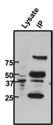
- Immunoprecipitation of CREB was performed on untreated 293T cells. The antigen:antbody complex was formed by binding 500 µg whole cell lysate with 2 µg of mouse monoclonal antibody recognizing CREB (Product # MA1-083) overnight on a rocking platform at 4°C. The immune-complex was then captured on 50 µL Protein A/G Plus Agarose (Product # 20423). Captured immune-complexes were then washed extensively and proteins eluted with 5X Reducing Sample Loading Dye (Product # 39000). Samples were then resolved on a 4-20% Tris-HCl polyacrylamide gel. Proteins were transferred to PVDF membrane and blocked with 5% Milk/TBS-0.1%Tween for at least 1 hour. Membranes were then probed with a mouse monoclonal antibody recognizing CREB (Product # MA1-083) at a dilution of 1:1000 overnight rotating at 4°C. Membranes were then washed in TBST and probed with a goat anti-mouse-HRP secondary antibody (Product # 32430) at a dilution of 1:20,000 for at least one hour. Membranes were washed and chemiluminescent detection was performed using Pierce Super Signal West Dura (Product # 34075).

Supportive validation
- Submitted by
- Invitrogen Antibodies (provider)
- Main image
- Experimental details
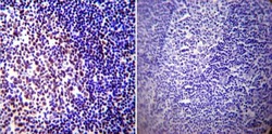
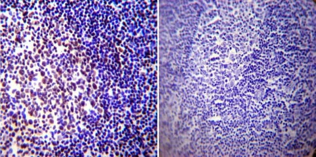
- Immunohistochemistry was performed on normal biopsies of deparaffinized Human tonsil tissue. To expose target proteins, heat induced antigen retrieval was performed using 10mM sodium citrate (pH6.0) buffer, microwaved for 8-15 minutes. Following antigen retrieval tissues were blocked in 3% BSA-PBS for 30 minutes at room temperature. Tissues were then probed at a dilution of 1:100 with a mouse monoclonal antibody recognizing CREB (Product # MA1-083) or without primary antibody (negative control) overnight at 4°C in a humidified chamber. Tissues were washed extensively with PBST and endogenous peroxidase activity was quenched with a peroxidase suppressor. Detection was performed using a biotin-conjugated secondary antibody and SA-HRP, followed by colorimetric detection using DAB. Tissues were counterstained with hematoxylin and prepped for mounting.
- Submitted by
- Invitrogen Antibodies (provider)
- Main image
- Experimental details
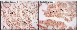
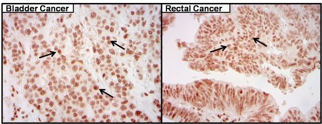
- Immunohistochemistry was performed on cancer biopsies of deparaffinized human bladder and rectal tissue. To expose target proteins high pressure heat induced antigen retrieval was performed using 10mM sodium citrate (pH6.0) buffer for 20 minutes. Following antigen retrieval endogenous peroxidase activity was quenched with 3% hydrogen peroxide for 10 minutes at room-temp. Tissues were then washed in PBS and blocked in 10% normal goat serum for 20 minutes at room temperature. Tissues were probed at a dilution of 1:200 with a mouse monoclonal antibody recognizing CREB (Product # MA1-083) overnight at 4°C in a humidified chamber. Tissues were washed extensively with PBS. Colorimetric detection was performed using metal enhanced DAB. Images are displayed at 40X magnification. Results demonstrate strong nuclear localization of CREB.
Supportive validation
- Submitted by
- Invitrogen Antibodies (provider)
- Main image
- Experimental details
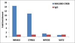
- Enrichment of endogenous CREB protein at specific gene loci using Anti-CREB Mouse Monoclonal Antibody: Chromatin Immunoprecipitation (ChIP) was performed using Anti-CREB Mouse Monoclonal Antibody (Product # MA1-083, 5 µg) on sheared chromatin from 2 million HeLa cells using the MAGnify ChIP system kit (Product # 49-2024). Normal Rabbit IgG was used as a negative IP control. The purified DNA was analyzed by 7500 Fast qPCR system (Product # 4351106) with optimized PCR primer pairs for the promoter of NR4A3, CYR61 gene used as positive control target, and the MYOD, SAT2, used as negative control target. Data is presented as fold enrichment of the antibody signal versus the negative control IgG using the comparative CT method.

- Submitted by
- Invitrogen Antibodies (provider)
- Main image

- Experimental details
- Enrichment of endogenous CREB protein at specific gene loci using Anti-CREB Mouse Monoclonal Antibody: Chromatin Immunoprecipitation (ChIP) was performed using Anti-CREB Mouse Monoclonal Antibody (Product # MA1-083, 5 µg) on sheared chromatin from 2 million HeLa cells using the MAGnify ChIP system kit (Product # 49-2024). Normal Rabbit IgG was used as a negative IP control. The purified DNA was analyzed by 7500 Fast qPCR system (Product # 4351106) with optimized PCR primer pairs for the promoter of NR4A3, CYR61 gene used as positive control target, and the MYOD, SAT2, used as negative control target. Data is presented as fold enrichment of the antibody signal versus the negative control IgG using the comparative CT method.

Supportive validation
- Submitted by
- Invitrogen Antibodies (provider)
- Main image
- Experimental details
- Immunoprecipitation of CREB was performed on untreated 293T cells. The antigen:antbody complex was formed by binding 500 µg whole cell lysate with 2 µg of mouse monoclonal antibody recognizing CREB (Product # MA1-083) overnight on a rocking platform at 4øC. The immune-complex was then captured on 50 µL Protein A/G Plus Agarose (Product # 20423). Captured immune-complexes were then washed extensively and proteins eluted with 5X Reducing Sample Loading Dye (Product # 39000). Samples were then resolved on a 4-20% Tris-HCl polyacrylamide gel. Proteins were transferred to PVDF membrane and blocked with 5% Milk/TBS-0.1%Tween for at least 1 hour. Membranes were then probed with a mouse monoclonal antibody recognizing CREB (Product # MA1-083) at a dilution of 1:1000 overnight rotating at 4øC. Membranes were then washed in TBST and probed with a goat anti-mouse-HRP secondary antibody (Product # 32430) at a dilution of 1:20,000 for at least one hour. Membranes were washed and chemiluminescent detection was performed using Pierce Super Signal West Dura (Product # 34075).

- Submitted by
- Invitrogen Antibodies (provider)
- Main image
- Experimental details
- NULL
- Submitted by
- Invitrogen Antibodies (provider)
- Main image
- Experimental details
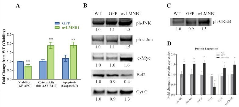
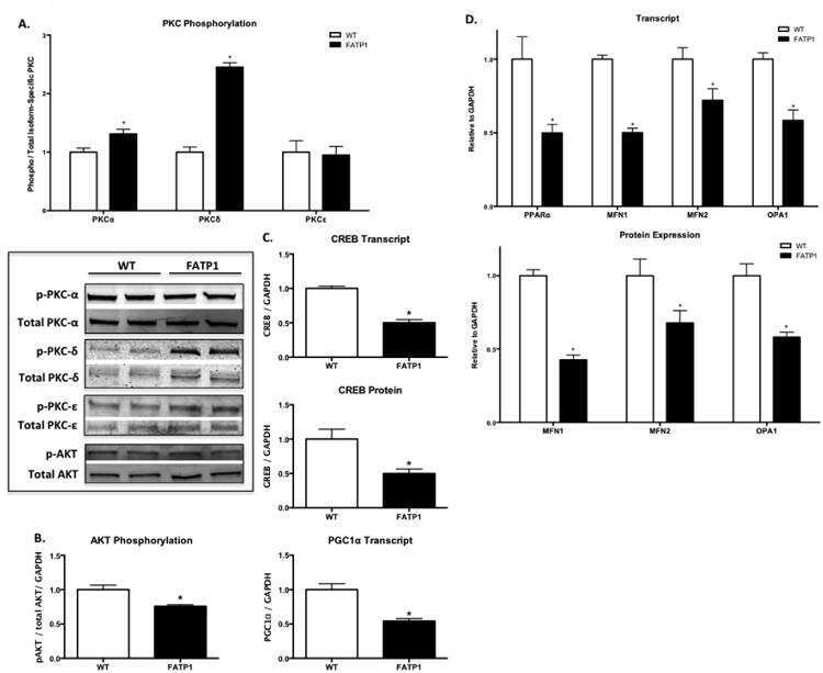
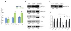
- Figure 4 Lamin B1 accumulation reduces cell viability and induces apoptosis signaling pathways: ( A ): Cell viability, cytotoxicity, and apoptosis of transduced cells (ovLMNB1 and GFP) were evaluated by using the ApoTox-Glo Triplex Assay and compared to wild type cells. Analyses from three independent experiments, with ** p < 0.01 vs. corresponding mock-transduced sample (GFP). ( B ): The activation of signaling pathways involved in apoptosis and the expression of downstream targets was evaluated in U87-MG cells 96 h after transduction. The expression or the phosphorylation of the denoted proteins were evaluated. ( C ): CREB phosphorylation was measured in wild type, mock-transduced, and Lamin B1--overexpressing cells. Samples were normalized by using total protein normalization vs. wild type cells. Results are representative of five independent experiments. ( D ): Quantitative analysis of protein expression; wild type cells (WT) were used as reference sample. ** denotes p < 0.01 vs. corresponding mock-transduced sample (GFP).