 Explore
 Validate
 Learn
LearnMA1-155
antibody from Invitrogen Antibodies
Targeting: CCNB1
CCNB
 Western blot
 Immunocytochemistry Immunoprecipitation Immunohistochemistry
 Flow cytometry Other assay
Antibody data
- Antibody Data
- Antigen structure
- References [3]
- Comments [0]
- Validations
- Immunocytochemistry [12]
- Immunoprecipitation [1]
- Immunohistochemistry [4]
- Other assay [1]
Submit
Validation data
Reference
Comment
Report error
- Product number
- MA1-155 - Provider product page

- Provider
- Invitrogen Antibodies
- Product name
- Cyclin B1 Monoclonal Antibody (V152)
- Antibody type
- Monoclonal
- Antigen
- Other
- Description
- MA1-155 detects Cyclin B1 in mammalian samples. MA1-155 has been successfully used in Immunohistochemistry (paraffin), Immunofluorescence, Immunoprecipitation, Flow Cytometry and Western Blot procedures. Immunoprecipitation and Western Blot analysis with MA1-155 show the accumulation of a prominent band at ~52 kDa in camptothecin and hydroxyurea treated cells. MA1-155 also detects additional unknown bands at ~35 and ~75 kDa. In Immunofluorescence applications, MA1-155 shows cyclin B1 co-localization with microtubules, whereas MA1-156 shows cyclin B2 staining consistent with the Golgi region. MA1-155 specifically detects Cyclin B1 in in whole cell lysates, but will also cross-react with purified recombinant human Cyclin B2 in Western Blot applications.
- Reactivity
- Human, Mouse, Rat, Hamster
- Host
- Mouse
- Isotype
- IgG
- Antibody clone number
- V152
- Vial size
- 100 μg
- Concentration
- 1 mg/mL
- Storage
- -20°C
Submitted references CircHACE1 functions as a competitive endogenous RNA to curb differentiated thyroid cancer progression by upregulating Tfcp2L1 through adsorbing miR-346.
Manipulation of the N-terminal sequence of the Borna disease virus X protein improves its mitochondrial targeting and neuroprotective potential.
Aurora kinase A is not involved in CPEB1 phosphorylation and cyclin B1 mRNA polyadenylation during meiotic maturation of porcine oocytes.
Li X, Yang S, Zhao C, Yang J, Li C, Shen W, Hu H, Zhang W, Yang S
Endocrine journal 2021 Aug 28;68(8):1011-1025
Endocrine journal 2021 Aug 28;68(8):1011-1025
Manipulation of the N-terminal sequence of the Borna disease virus X protein improves its mitochondrial targeting and neuroprotective potential.
Ferré CA, Davezac N, Thouard A, Peyrin JM, Belenguer P, Miquel MC, Gonzalez-Dunia D, Szelechowski M
FASEB journal : official publication of the Federation of American Societies for Experimental Biology 2016 Apr;30(4):1523-33
FASEB journal : official publication of the Federation of American Societies for Experimental Biology 2016 Apr;30(4):1523-33
Aurora kinase A is not involved in CPEB1 phosphorylation and cyclin B1 mRNA polyadenylation during meiotic maturation of porcine oocytes.
Komrskova P, Susor A, Malik R, Prochazkova B, Liskova L, Supolikova J, Hladky S, Kubelka M
PloS one 2014;9(7):e101222
PloS one 2014;9(7):e101222
No comments: Submit comment
Supportive validation
- Submitted by
- Invitrogen Antibodies (provider)
- Main image

- Experimental details
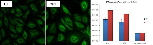
- Immunofluorescent analysis of Cyclin B1 (green) in HeLa cells left untreated (UT, left panel) or treated with 100nM camptothecin (CPT, middle panel). Formalin-fixed cells were permeabilized with 0.1% Triton X-100 in TBS and blocked with 1% Blocker BSA (Product # 37525) for 15 minutes at room temperature. Cells were probed with a Cyclin B1 monoclonal antibody (Product # MA1-155) at a dilution of 1:100 for at least 1 hour at room temperature, washed with PBS, and incubated with a DyLight 488-conjugated goat anti-mouse IgG secondary antibody (Product # 35502). Images were taken on a Thermo Scientific ToxInsight Instrument at 20X magnification. Accumulation of cytoplasmic/perinuclear Cyclin B1 in response to camptothecin treatment (right panel) was analyzed for 1:50 and 1:100 dilutions of MA1-155 displayed as RFU.

- Submitted by
- Invitrogen Antibodies (provider)
- Main image
- Experimental details
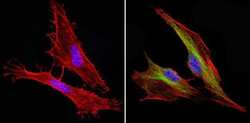
- Immunofluorescent analysis of Cyclin B1 (green) showing staining in the in the cytoplasm and nucleus of C2C12 cells (right) compared to a negative control without primary antibody (left). Formalin-fixed cells were permeabilized with 0.1% Triton X-100 in TBS for 5-10 minutes and blocked with 3% BSA-PBS for 30 minutes at room temperature. Cells were probed with a Cyclin B1 monoclonal antibody (Product # MA1-155) in 3% BSA-PBS at a dilution of 1:150 and incubated overnight at 4ºC in a humidified chamber. Cells were washed with PBST and incubated with a DyLight-conjugated secondary antibody in PBS at room temperature in the dark. F-actin (red) was stained with a fluorescent red phalloidin and nuclei (blue) were stained with Hoechst or DAPI. Images were taken at a magnification of 60x.
- Submitted by
- Invitrogen Antibodies (provider)
- Main image
- Experimental details
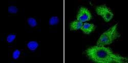
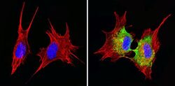
- Immunofluorescent analysis of Cyclin B1 (green) showing staining in the in the cytoplasm and nucleus of Hela cells (right) compared to a negative control without primary antibody (left). Formalin-fixed cells were permeabilized with 0.1% Triton X-100 in TBS for 5-10 minutes and blocked with 3% BSA-PBS for 30 minutes at room temperature. Cells were probed with a Cyclin B1 monoclonal antibody (Product # MA1-155) in 3% BSA-PBS at a dilution of 1:100 and incubated overnight at 4ºC in a humidified chamber. Cells were washed with PBST and incubated with a DyLight-conjugated secondary antibody in PBS at room temperature in the dark. F-actin (red) was stained with a fluorescent red phalloidin and nuclei (blue) were stained with Hoechst or DAPI. Images were taken at a magnification of 60x.
- Submitted by
- Invitrogen Antibodies (provider)
- Main image
- Experimental details
- Immunofluorescent analysis of Cyclin B1 (green) showing staining in the in the cytoplasm and nucleus of HepG2 cells (right) compared to a negative control without primary antibody (left). Formalin-fixed cells were permeabilized with 0.1% Triton X-100 in TBS for 5-10 minutes and blocked with 3% BSA-PBS for 30 minutes at room temperature. Cells were probed with a Cyclin B1 monoclonal antibody (Product # MA1-155) in 3% BSA-PBS at a dilution of 1:150 and incubated overnight at 4ºC in a humidified chamber. Cells were washed with PBST and incubated with a DyLight-conjugated secondary antibody in PBS at room temperature in the dark. Nuclei (blue) were stained with Hoechst or DAPI. Images were taken at a magnification of 60x.

- Submitted by
- Invitrogen Antibodies (provider)
- Main image

- Experimental details
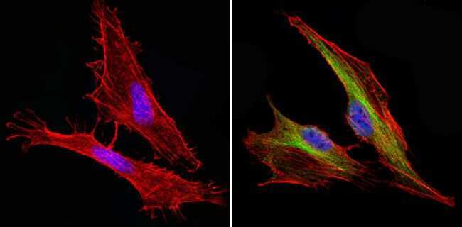
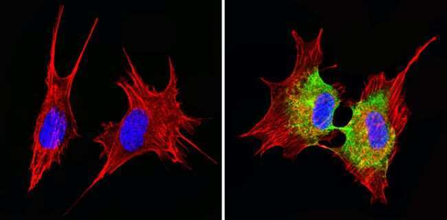
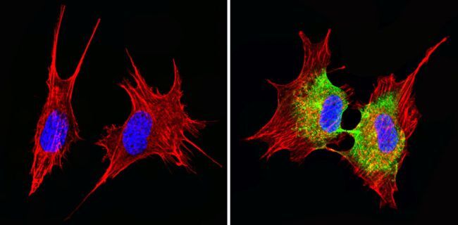
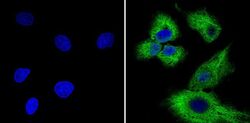
- Immunofluorescent analysis of Cyclin B1 (green) showing staining in the in the cytoplasm and nucleus of NIH-3T3 cells (right) compared to a negative control without primary antibody (left). Formalin-fixed cells were permeabilized with 0.1% Triton X-100 in TBS for 5-10 minutes and blocked with 3% BSA-PBS for 30 minutes at room temperature. Cells were probed with a Cyclin B1 monoclonal antibody (Product # MA1-155) in 3% BSA-PBS at a dilution of 1:150 and incubated overnight at 4ºC in a humidified chamber. Cells were washed with PBST and incubated with a DyLight-conjugated secondary antibody in PBS at room temperature in the dark. F-actin (red) was stained with a fluorescent red phalloidin and nuclei (blue) were stained with Hoechst or DAPI. Images were taken at a magnification of 60x.
- Submitted by
- Invitrogen Antibodies (provider)
- Main image
- Experimental details
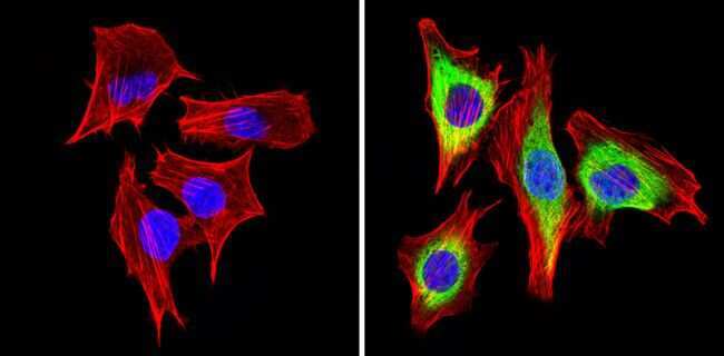
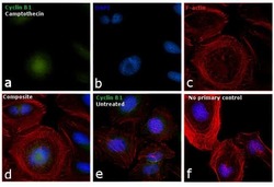
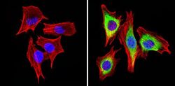
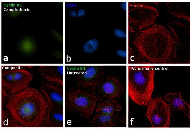
- Immunofluorescence analysis of Cyclin B1 was performed using 70% confluent log phase HeLa cells treated with Camptothecin (100nM, 16 hrs). The cells were fixed with 4% paraformaldehyde for 10 minutes, permeabilized with 0.1% Triton™ X-100 for 15 minutes, and blocked with 1% BSA for 1 hour at room temperature. The cells were labeled with Cyclin B1 Monoclonal Antibody (Product # MA1-155) at 1:50 dilution in 0.1% BSA, incubated at 4 degree Celsius overnight and then labeled with Goat anti-Mouse IgG (H+L) Superclonal™ Secondary Antibody, Alexa Fluor® 488 conjugate (Product # A28175) at a dilution of 1:2000 for 45 minutes at room temperature (Panel a: green). Nuclei (Panel b: blue) were stained with ProLong™ Diamond Antifade Mountant with DAPI (Product # P36962). F-actin (Panel c: red) was stained with Rhodamine Phalloidin (Product # R415, 1:300). Panel d represents the merged image showing accumulation of Cyclin A2 in the nucleus upon treatment. Panel e represents the control cells showing cytoplasmic staining. Panel f represents control cells with no primary antibody to assess background. The images were captured at 60X magnification.
- Submitted by
- Invitrogen Antibodies (provider)
- Main image

- Experimental details
- Immunofluorescent analysis of Cyclin B1 (green) in HeLa cells left untreated (UT, left panel) or treated with 100nM camptothecin (CPT, middle panel). Formalin-fixed cells were permeabilized with 0.1% Triton X-100 in TBS and blocked with 1% Blocker BSA (Product # 37525) for 15 minutes at room temperature. Cells were probed with a Cyclin B1 monoclonal antibody (Product # MA1-155) at a dilution of 1:100 for at least 1 hour at room temperature, washed with PBS, and incubated with a DyLight 488-conjugated goat anti-mouse IgG secondary antibody (Product # 35502). Images were taken on a Thermo Scientific ToxInsight Instrument at 20X magnification. Accumulation of cytoplasmic/perinuclear Cyclin B1 in response to camptothecin treatment (right panel) was analyzed for 1:50 and 1:100 dilutions of MA1-155 displayed as RFU.
- Submitted by
- Invitrogen Antibodies (provider)
- Main image

- Experimental details
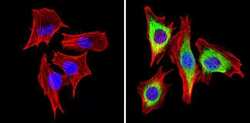
- Immunofluorescent analysis of Cyclin B1 (green) showing staining in the in the cytoplasm and nucleus of Hela cells (right) compared to a negative control without primary antibody (left). Formalin-fixed cells were permeabilized with 0.1% Triton X-100 in TBS for 5-10 minutes and blocked with 3% BSA-PBS for 30 minutes at room temperature. Cells were probed with a Cyclin B1 monoclonal antibody (Product # MA1-155) in 3% BSA-PBS at a dilution of 1:100 and incubated overnight at 4ºC in a humidified chamber. Cells were washed with PBST and incubated with a DyLight-conjugated secondary antibody in PBS at room temperature in the dark. F-actin (red) was stained with a fluorescent red phalloidin and nuclei (blue) were stained with Hoechst or DAPI. Images were taken at a magnification of 60x.

- Submitted by
- Invitrogen Antibodies (provider)
- Main image
- Experimental details
- Immunofluorescent analysis of Cyclin B1 (green) showing staining in the in the cytoplasm and nucleus of C2C12 cells (right) compared to a negative control without primary antibody (left). Formalin-fixed cells were permeabilized with 0.1% Triton X-100 in TBS for 5-10 minutes and blocked with 3% BSA-PBS for 30 minutes at room temperature. Cells were probed with a Cyclin B1 monoclonal antibody (Product # MA1-155) in 3% BSA-PBS at a dilution of 1:150 and incubated overnight at 4ºC in a humidified chamber. Cells were washed with PBST and incubated with a DyLight-conjugated secondary antibody in PBS at room temperature in the dark. F-actin (red) was stained with a fluorescent red phalloidin and nuclei (blue) were stained with Hoechst or DAPI. Images were taken at a magnification of 60x.

- Submitted by
- Invitrogen Antibodies (provider)
- Main image
- Experimental details
- Immunofluorescent analysis of Cyclin B1 (green) showing staining in the in the cytoplasm and nucleus of NIH-3T3 cells (right) compared to a negative control without primary antibody (left). Formalin-fixed cells were permeabilized with 0.1% Triton X-100 in TBS for 5-10 minutes and blocked with 3% BSA-PBS for 30 minutes at room temperature. Cells were probed with a Cyclin B1 monoclonal antibody (Product # MA1-155) in 3% BSA-PBS at a dilution of 1:150 and incubated overnight at 4ºC in a humidified chamber. Cells were washed with PBST and incubated with a DyLight-conjugated secondary antibody in PBS at room temperature in the dark. F-actin (red) was stained with a fluorescent red phalloidin and nuclei (blue) were stained with Hoechst or DAPI. Images were taken at a magnification of 60x.
- Submitted by
- Invitrogen Antibodies (provider)
- Main image
- Experimental details
- Immunofluorescent analysis of Cyclin B1 (green) showing staining in the in the cytoplasm and nucleus of HepG2 cells (right) compared to a negative control without primary antibody (left). Formalin-fixed cells were permeabilized with 0.1% Triton X-100 in TBS for 5-10 minutes and blocked with 3% BSA-PBS for 30 minutes at room temperature. Cells were probed with a Cyclin B1 monoclonal antibody (Product # MA1-155) in 3% BSA-PBS at a dilution of 1:150 and incubated overnight at 4ºC in a humidified chamber. Cells were washed with PBST and incubated with a DyLight-conjugated secondary antibody in PBS at room temperature in the dark. Nuclei (blue) were stained with Hoechst or DAPI. Images were taken at a magnification of 60x.

- Submitted by
- Invitrogen Antibodies (provider)
- Main image

- Experimental details
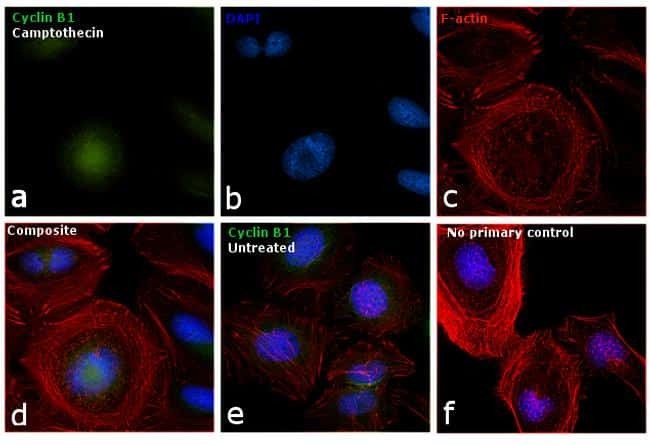
- Immunofluorescence analysis of Cyclin B1 was performed using 70% confluent log phase HeLa cells treated with Camptothecin (100nM, 16 hrs). The cells were fixed with 4% paraformaldehyde for 10 minutes, permeabilized with 0.1% Triton™ X-100 for 15 minutes, and blocked with 1% BSA for 1 hour at room temperature. The cells were labeled with Cyclin B1 Monoclonal Antibody (Product # MA1-155) at 1:50 dilution in 0.1% BSA, incubated at 4 degree Celsius overnight and then labeled with Goat anti-Mouse IgG (H+L) Superclonal™ Secondary Antibody, Alexa Fluor® 488 conjugate (Product # A28175) at a dilution of 1:2000 for 45 minutes at room temperature (Panel a: green). Nuclei (Panel b: blue) were stained with ProLong™ Diamond Antifade Mountant with DAPI (Product # P36962). F-actin (Panel c: red) was stained with Rhodamine Phalloidin (Product # R415, 1:300). Panel d represents the merged image showing accumulation of Cyclin A2 in the nucleus upon treatment. Panel e represents the control cells showing cytoplasmic staining. Panel f represents control cells with no primary antibody to assess background. The images were captured at 60X magnification.
Supportive validation
- Submitted by
- Invitrogen Antibodies (provider)
- Main image

- Experimental details
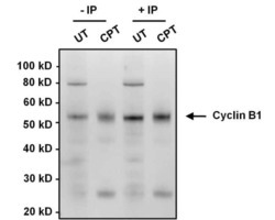
- Immunoprecipitation analysis of Cyclin B1 was performed on HeLa cell lysates, from cells left untreated (UT) or treated with 100 nM camptothecin (CPT). Antigen-antibody complexes were formed by incubating 375 µg of lysate with 1 µg of a Cyclin B1 monoclonal antibody (Product # MA1-155) overnight on an end-over-end rotator at 4°C. The immune complexes were captured on 100 µL Protein A/G Plus Agarose (Product # 20423), washed extensively, and eluted with 5X Lane Marker Reducing Sample Buffer (Product # 39000). Samples, including control input cell lysates (-IP lanes), were resolved on a 4-12% Bis-Tris polyacrylamide gel, transferred to a nitrocellulose membrane and blocked with 5% BSA/TBST for 1 hour. The membrane was probed with a Cyclin B1 monoclonal antibody (Product # MA1-155) at a dilution of 1:1000 overnight rotating at 4°C, washed in TBST, and probed with Clean-Blot IP Detection Reagent (Product # 21230). Chemiluminescent detection was performed using SuperSignal West Dura (Product # 34076).

Supportive validation
- Submitted by
- Invitrogen Antibodies (provider)
- Main image
- Experimental details
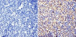
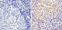
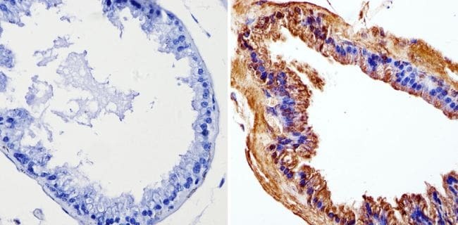
- Immunohistochemistry analysis of Cyclin B1 showing staining in the nucleus and cytoplasm of paraffin-embedded mouse lymph node tissue (right) compared with a negative control without primary antibody (left). To expose target proteins, antigen retrieval was performed using 10mM sodium citrate (pH 6.0), microwaved for 8-15 min. Following antigen retrieval, tissues were blocked in 3% H2O2-methanol for 15 min at room temperature, washed with ddH2O and PBS, and then probed with a Cyclin B1 monoclonal antibody (Product # MA1-155) diluted in 3% BSA-PBS at a dilution of 1:20 overnight at 4°C in a humidified chamber. Tissues were washed extensively in PBST and detection was performed using an HRP-conjugated secondary antibody followed by colorimetric detection using a DAB kit. Tissues were counterstained with hematoxylin and dehydrated with ethanol and xylene to prep for mounting.
- Submitted by
- Invitrogen Antibodies (provider)
- Main image
- Experimental details
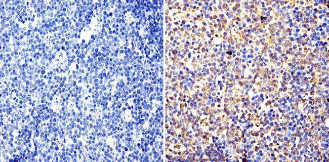
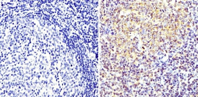
- Immunohistochemistry analysis of Cyclin B1 showing staining in the nucleus and cytoplasm of paraffin-embedded human tonsil tissue (right) compared with a negative control without primary antibody (left). To expose target proteins, antigen retrieval was performed using 10mM sodium citrate (pH 6.0), microwaved for 8-15 min. Following antigen retrieval, tissues were blocked in 3% H2O2-methanol for 15 min at room temperature, washed with ddH2O and PBS, and then probed with a Cyclin B1 monoclonal antibody (Product # MA1-155) diluted in 3% BSA-PBS at a dilution of 1:200 overnight at 4°C in a humidified chamber. Tissues were washed extensively in PBST and detection was performed using an HRP-conjugated secondary antibody followed by colorimetric detection using a DAB kit. Tissues were counterstained with hematoxylin and dehydrated with ethanol and xylene to prep for mounting.
- Submitted by
- Invitrogen Antibodies (provider)
- Main image
- Experimental details
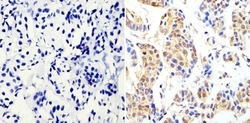
- Immunohistochemistry analysis of Cyclin B1 showing staining in the nucleus and cytoplasm of paraffin-embedded human breast carcinoma (right) compared with a negative control without primary antibody (left). To expose target proteins, antigen retrieval was performed using 10mM sodium citrate (pH 6.0), microwaved for 8-15 min. Following antigen retrieval, tissues were blocked in 3% H2O2-methanol for 15 min at room temperature, washed with ddH2O and PBS, and then probed with a Cyclin B1 monoclonal antibody (Product # MA1-155) diluted in 3% BSA-PBS at a dilution of 1:200 overnight at 4°C in a humidified chamber. Tissues were washed extensively in PBST and detection was performed using an HRP-conjugated secondary antibody followed by colorimetric detection using a DAB kit. Tissues were counterstained with hematoxylin and dehydrated with ethanol and xylene to prep for mounting.
- Submitted by
- Invitrogen Antibodies (provider)
- Main image

- Experimental details
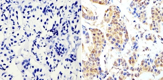
- Immunohistochemistry analysis of Cyclin B1 showing staining in the nucleus and cytoplasm of paraffin-embedded mouse prostate tissue (right) compared with a negative control without primary antibody (left). To expose target proteins, antigen retrieval was performed using 10mM sodium citrate (pH 6.0), microwaved for 8-15 min. Following antigen retrieval, tissues were blocked in 3% H2O2-methanol for 15 min at room temperature, washed with ddH2O and PBS, and then probed with a Cyclin B1 monoclonal antibody (Product # MA1-155) diluted in 3% BSA-PBS at a dilution of 1:100 overnight at 4°C in a humidified chamber. Tissues were washed extensively in PBST and detection was performed using an HRP-conjugated secondary antibody followed by colorimetric detection using a DAB kit. Tissues were counterstained with hematoxylin and dehydrated with ethanol and xylene to prep for mounting.
Supportive validation
- Submitted by
- Invitrogen Antibodies (provider)
- Main image
- Experimental details
- Immunoprecipitation analysis of Cyclin B1 was performed on HeLa cell lysates, from cells left untreated (UT) or treated with 100 nM camptothecin (CPT). Antigen-antibody complexes were formed by incubating 375 µg of lysate with 1 µg of a Cyclin B1 monoclonal antibody (Product # MA1-155) overnight on an end-over-end rotator at 4øC. The immune complexes were captured on 100 µL Protein A/G Plus Agarose (Product # 20423), washed extensively, and eluted with 5X Lane Marker Reducing Sample Buffer (Product # 39000). Samples, including control input cell lysates (-IP lanes), were resolved on a 4-12% Bis-Tris polyacrylamide gel, transferred to a nitrocellulose membrane and blocked with 5% BSA/TBST for 1 hour. The membrane was probed with a Cyclin B1 monoclonal antibody (Product # MA1-155) at a dilution of 1:1000 overnight rotating at 4øC, washed in TBST, and probed with Clean-Blot IP Detection Reagent (Product # 21230). Chemiluminescent detection was performed using SuperSignal West Dura (Product # 34076).
