 Explore
 Validate
 Learn
Learn Western blot
Antibody data
- Antibody Data
- Antigen structure
- References [2]
- Comments [0]
- Validations
- Western blot [2]
- Immunohistochemistry [1]
Submit
Validation data
Reference
Comment
Report error
- Product number
- AF6000 - Provider product page

- Provider
- Novus Biologicals
- Product name
- Goat Polyclonal Cyclin B1 Antibody
- Antibody type
- Polyclonal
- Description
- Antigen Affinity-purified. Detects human and mouse Cyclin B1 in direct ELISAs and Western blots.
- Reactivity
- Human, Mouse
- Host
- Goat
- Conjugate
- Unconjugated
- Isotype
- IgG
- Vial size
- 100 ug
- Concentration
- LYOPH
- Storage
- Use a manual defrost freezer and avoid repeated freeze-thaw cycles. 12 months from date of receipt, -20 to -70 degreesC as supplied. 1 month, 2 to 8 degreesC under sterile conditions after reconstitution. 6 months, -20 to -70 degreesC under sterile conditions after reconstitution.
Submitted references Enhanced Expression of the Key Mitosis Regulator Cyclin B1 Is Mediated by PDZ-Binding Kinase in Islets of Pregnant Mice.
Jab1 regulates Schwann cell proliferation and axonal sorting through p27.
Uesato T, Ogihara T, Hara A, Iida H, Miyatsuka T, Fujitani Y, Takeda S, Watada H
Journal of the Endocrine Society 2018 Mar 1;2(3):207-219
Journal of the Endocrine Society 2018 Mar 1;2(3):207-219
Jab1 regulates Schwann cell proliferation and axonal sorting through p27.
Porrello E, Rivellini C, Dina G, Triolo D, Del Carro U, Ungaro D, Panattoni M, Feltri ML, Wrabetz L, Pardi R, Quattrini A, Previtali SC
The Journal of experimental medicine 2014 Jan 13;211(1):29-43
The Journal of experimental medicine 2014 Jan 13;211(1):29-43
No comments: Submit comment
Supportive validation
- Submitted by
- Novus Biologicals (provider)
- Main image

- Experimental details
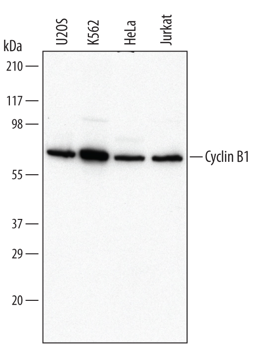
- Detection of Human Cyclin B1 by Western Blot. Western blot shows lysates of U2OS human osteosarcoma cell line, K562 human chronic myelogenous leukemia cell line, HeLa human cervical epithelial carcinoma cell line, and Jurkat human acute T cell leukemia cell line. PVDF Membrane was probed with 0.2 µg/mL of Goat Anti-Human/Mouse Cyclin B1 Antigen Affinity-purified Polyclonal Antibody (Catalog # AF6000) followed by HRP-conjugated Anti-Goat IgG Secondary Antibody (Catalog # HAF017). A specific band was detected for Cyclin B1 at approximately 62 kDa (as indicated). This experiment was conducted under reducing conditions and using Immunoblot Buffer Group 1.
- Submitted by
- Novus Biologicals (provider)
- Main image

- Experimental details
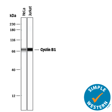
- Detection of Human Cyclin B1 by Simple WesternTM. Simple Western lane view shows lysates of HeLa human cervical epithelial carcinoma cell line and Jurkat human acute T cell leukemia cell line, loaded at 0.2 mg/mL. A specific band was detected for Cyclin B1 at approximately 67 kDa (as indicated) using 2 µg/mL of Goat Anti-Human/Mouse Cyclin B1 Antigen Affinity-purified Polyclonal Antibody (Catalog # AF6000) followed by 1:50 dilution of HRP-conjugated Anti-Goat IgG Secondary Antibody (Catalog # HAF109). This experiment was conducted under reducing conditions and using the 12-230 kDa separation system.
Supportive validation
- Submitted by
- Novus Biologicals (provider)
- Main image
- Experimental details
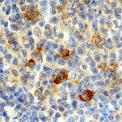
- Cyclin B1 in Human Lymphoma. Cyclin B1 was detected in immersion fixed paraffin-embedded sections of human lymphoma using Goat Anti-Human/Mouse Cyclin B1 Antigen Affinity-purified Polyclonal Antibody (Catalog # AF6000) at 10 µg/mL overnight at 4 °C. Before incubation with the primary antibody, tissue was subjected to heat-induced epitope retrieval using Antigen Retrieval Reagent-Basic (Catalog # CTS013). Tissue was stained using the Anti-Goat HRP-DAB Cell & Tissue Staining Kit (brown; Catalog # CTS008) and counter-stained with hematoxylin (blue). Specific staining was localized to cytoplasm in lymphocytes. View our protocol for Chromogenic IHC Staining of Paraffin-embedded Tissue Sections.
