 Explore
 Validate
 Learn
Learn Western blot
Antibody data
- Antibody Data
- Antigen structure
- References [2]
- Comments [0]
- Validations
- Western blot [2]
- Immunohistochemistry [1]
Submit
Validation data
Reference
Comment
Report error
- Product number
- PAB9943 - Provider product page

- Provider
- Abnova Corporation
- Proper citation
- Abnova Corporation Cat#PAB9943, RRID:AB_1672215
- Product name
- CCNB1 polyclonal antibody
- Antibody type
- Polyclonal
- Description
- Rabbit polyclonal antibody raised against full length recombinant CCNB1.
- Storage
- Store at 4°C. For long term storage store at -20°C.Aliquot to avoid repeated freezing and thawing.
Submitted references Human cyclin A is adenovirus E1A-associated protein p60 and behaves differently from cyclin B.
A 60 kd cdc2-associated polypeptide complexes with the E1A proteins in adenovirus-infected cells.
Pines J, Hunter T
Nature 1990 Aug 23;346(6286):760-3
Nature 1990 Aug 23;346(6286):760-3
A 60 kd cdc2-associated polypeptide complexes with the E1A proteins in adenovirus-infected cells.
Giordano A, Whyte P, Harlow E, Franza BR Jr, Beach D, Draetta G
Cell 1989 Sep 8;58(5):981-90
Cell 1989 Sep 8;58(5):981-90
No comments: Submit comment
Supportive validation
- Submitted by
- Abnova Corporation (provider)
- Main image
- Experimental details
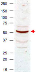
- Western blot analysis using CCNB1 polyclonal antibody (Cat # PAB9943) shows detection of CCNB1 present in asynchronous HeLa cell lysates.Comparison to a molecular weight marker indicates a band of ~55 KDa corresponding to human CCNB1 (arrowhead).Approximately 50 ug of lysate was loaded on to a 7% SDS-PAGE gel for separation.After transfer to nitrocellulose, the blot was incubated with a 1 : 500 dilution of the antibody for 1 h at room temperature.Detection occurred using a 1 : 10,000 of HRP conjugated Goat-a-Rabbit IgG.Personal communication, Luca D'Agostino, Temple University, Philadelphia, PA.

- Submitted by
- Abnova Corporation (provider)
- Main image
- Experimental details
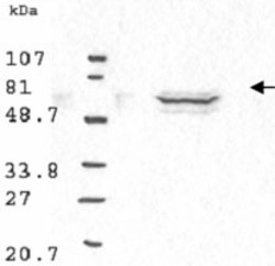
- Western blot analysis using CCNB1 polyclonal antibody (Cat # PAB9943) antibody shows detection of human CCNB1 present in asynchronous HN30 cell lysates.HN30 cells are from head and neck cancer tumors that over express CCNB1 and D1.Comparison to a molecular weight marker indicates a band of ~62 KDa corresponding to the expected molecular weight for the protein (arrowhead).The blot was incubated with a 1 : 500 dilution of the antibody for 1 h at room temperature.Detection occurred using a 1 : 10,000 of HRP conjugated Goat-a-Rabbit IgG and chemiluminescence reagent with a 1-min exposure time.Personal communication, Luca Cote, Temple University, Philadelphia, PA.

Supportive validation
- Submitted by
- Abnova Corporation (provider)
- Main image
- Experimental details
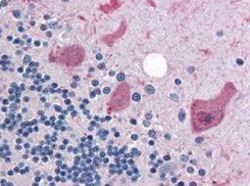
- Immunohistochemical staining with CCNB1 polyclonal antibody (Cat # PAB9943) was diluted 1 : 500 to detect CCNB1 in human brain cerebellum tissue.Tissue was formalin fixed and paraffin embedded.No pre-treatment of sample was required.The image shows the localization of antibody as the precipitated red signal, with a hematoxylin purple nuclear counter stain.
- Validation comment
- Immunohistochemistry (Formalin/PFA-fixed paraffin-embedded sections)
