 Explore
 Validate
 Learn
Learn Western blot
 Immunocytochemistry
Antibody data
- Antibody Data
- Antigen structure
- References [9]
- Comments [0]
- Validations
- Immunocytochemistry [7]
- Immunoprecipitation [1]
- Immunohistochemistry [1]
- Other assay [4]
Submit
Validation data
Reference
Comment
Report error
- Product number
- MA5-14327 - Provider product page

- Provider
- Invitrogen Antibodies
- Product name
- Cyclin B1 Monoclonal Antibody (GNS11)
- Antibody type
- Monoclonal
- Antigen
- Other
- Description
- MA5-14327 targets Cyclin B1 in WB, ICC/IF, IHC (P), and IP applications and shows reactivity with Hamster, Human, mouse, and Rat samples. This antibody is not recommended for NIH-3T3 cells in Western blot applications. The MA5-14327 immunogen is human cyclin B1 protein. This antibody was orginally validated as part of a Thermo Scientific Cellomics High Content Screening Kit. The antibody sold separately may have slightly different performance and may need to be further optimized for the best results.
- Reactivity
- Human, Mouse, Rat, Hamster
- Host
- Mouse
- Isotype
- IgG
- Antibody clone number
- GNS11
- Vial size
- 500 μL
- Concentration
- 0.2 mg/mL
- Storage
- 4°C
Submitted references A Hybrid Chalcone Combining the Trimethoxyphenyl and Isatinyl Groups Targets Multiple Oncogenic Proteins and Pathways in Hepatocellular Carcinoma Cells.
Proteomic Interaction Patterns between Human Cyclins, the Cyclin-Dependent Kinase Ortholog pUL97 and Additional Cytomegalovirus Proteins.
The Interaction between Cyclin B1 and Cytomegalovirus Protein Kinase pUL97 is Determined by an Active Kinase Domain.
MYC expression and distribution in normal mature lymphoid cells.
Mapping differentiation kinetics in the mouse retina reveals an extensive period of cell cycle protein expression in post-mitotic newborn neurons.
Cyclin A and cyclin B1 overexpression in differentiated thyroid carcinoma.
Immunohistochemical analysis of non-small cell lung cancer: correlation with clinical parameters and prognosis.
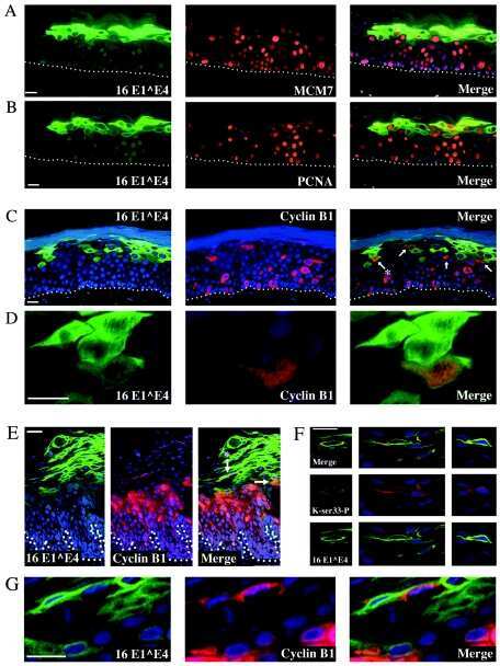
Human papillomavirus type 16 E1 E4-induced G2 arrest is associated with cytoplasmic retention of active Cdk1/cyclin B1 complexes.
Mitotic cell cycle proteins increase in podocytes despite lack of proliferation.
Cao L, Zhang L, Zhao X, Zhang Y
PloS one 2016;11(8):e0161025
PloS one 2016;11(8):e0161025
Proteomic Interaction Patterns between Human Cyclins, the Cyclin-Dependent Kinase Ortholog pUL97 and Additional Cytomegalovirus Proteins.
Steingruber M, Kraut A, Socher E, Sticht H, Reichel A, Stamminger T, Amin B, Couté Y, Hutterer C, Marschall M
Viruses 2016 Aug 18;8(8)
Viruses 2016 Aug 18;8(8)
The Interaction between Cyclin B1 and Cytomegalovirus Protein Kinase pUL97 is Determined by an Active Kinase Domain.
Steingruber M, Socher E, Hutterer C, Webel R, Bergbrede T, Lenac T, Sticht H, Marschall M
Viruses 2015 Aug 11;7(8):4582-601
Viruses 2015 Aug 11;7(8):4582-601
MYC expression and distribution in normal mature lymphoid cells.
Cattoretti G
The Journal of pathology 2013 Feb;229(3):430-40
The Journal of pathology 2013 Feb;229(3):430-40
Mapping differentiation kinetics in the mouse retina reveals an extensive period of cell cycle protein expression in post-mitotic newborn neurons.
Pacal M, Bremner R
Developmental dynamics : an official publication of the American Association of Anatomists 2012 Oct;241(10):1525-44
Developmental dynamics : an official publication of the American Association of Anatomists 2012 Oct;241(10):1525-44
Cyclin A and cyclin B1 overexpression in differentiated thyroid carcinoma.
Nar A, Ozen O, Tutuncu NB, Demirhan B
Medical oncology (Northwood, London, England) 2012 Mar;29(1):294-300
Medical oncology (Northwood, London, England) 2012 Mar;29(1):294-300
Immunohistochemical analysis of non-small cell lung cancer: correlation with clinical parameters and prognosis.
Yoo J, Jung JH, Lee MA, Seo KJ, Shim BY, Kim SH, Cho DG, Ahn MI, Kim CH, Cho KD, Kang SJ, Kim HK
Journal of Korean medical science 2007 Apr;22(2):318-25
Journal of Korean medical science 2007 Apr;22(2):318-25
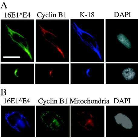
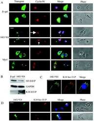
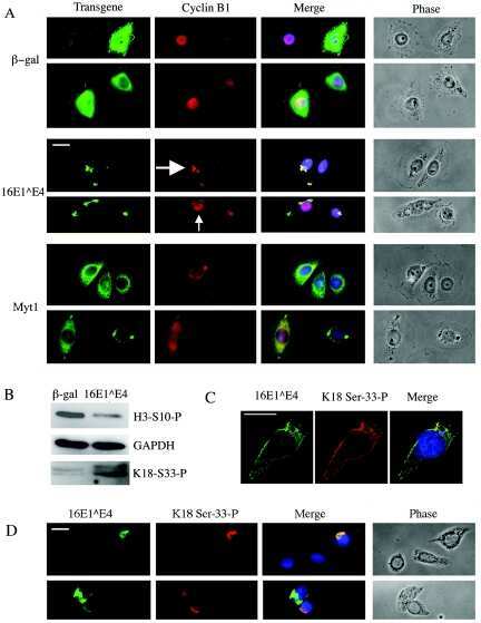
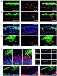
Human papillomavirus type 16 E1 E4-induced G2 arrest is associated with cytoplasmic retention of active Cdk1/cyclin B1 complexes.
Davy CE, Jackson DJ, Raj K, Peh WL, Southern SA, Das P, Sorathia R, Laskey P, Middleton K, Nakahara T, Wang Q, Masterson PJ, Lambert PF, Cuthill S, Millar JB, Doorbar J
Journal of virology 2005 Apr;79(7):3998-4011
Journal of virology 2005 Apr;79(7):3998-4011
Mitotic cell cycle proteins increase in podocytes despite lack of proliferation.
Petermann AT, Pippin J, Hiromura K, Monkawa T, Durvasula R, Couser WG, Kopp J, Shankland SJ
Kidney international 2003 Jan;63(1):113-22
Kidney international 2003 Jan;63(1):113-22
No comments: Submit comment
Supportive validation
- Submitted by
- Invitrogen Antibodies (provider)
- Main image
- Experimental details
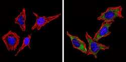
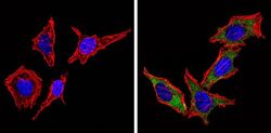
- Immunofluorescent analysis of Cyclin B1 (green) showing staining in the cytoplasm of Hela cells (right) compared to a negative control without primary antibody (left). Formalin-fixed cells were permeabilized with 0.1% Triton X-100 in TBS for 5-10 minutes and blocked with 3% BSA-PBS for 30 minutes at room temperature. Cells were probed with a Cyclin B1 monoclonal antibody (Product # MA5-14327) in 3% BSA-PBS at a dilution of 1:20 and incubated overnight at 4 ºC in a humidified chamber. Cells were washed with PBST and incubated with a DyLight-conjugated secondary antibody in PBS at room temperature in the dark. F-actin (red) was stained with a fluorescent red phalloidin and nuclei (blue) were stained with Hoechst or DAPI. Images were taken at a magnification of 60x.
- Submitted by
- Invitrogen Antibodies (provider)
- Main image

- Experimental details
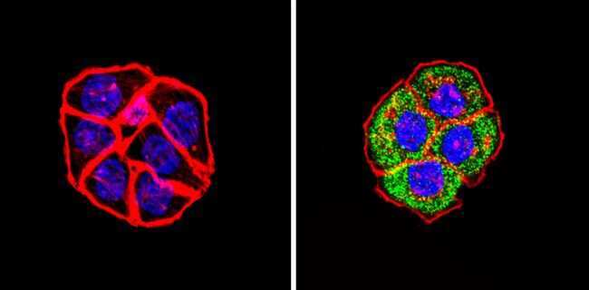
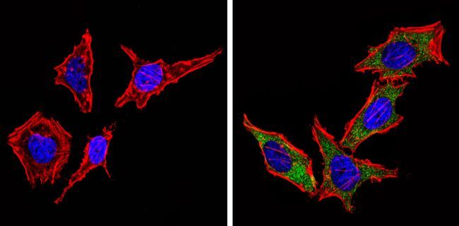
- Immunofluorescent analysis of Cyclin B1 (green) showing staining in the cytoplasm of HT29 cells (right) compared to a negative control without primary antibody (left). Formalin-fixed cells were permeabilized with 0.1% Triton X-100 in TBS for 5-10 minutes and blocked with 3% BSA-PBS for 30 minutes at room temperature. Cells were probed with a Cyclin B1 monoclonal antibody (Product # MA5-14327) in 3% BSA-PBS at a dilution of 1:20 and incubated overnight at 4 ºC in a humidified chamber. Cells were washed with PBST and incubated with a DyLight-conjugated secondary antibody in PBS at room temperature in the dark. F-actin (red) was stained with a fluorescent red phalloidin and nuclei (blue) were stained with Hoechst or DAPI. Images were taken at a magnification of 60x.
- Submitted by
- Invitrogen Antibodies (provider)
- Main image
- Experimental details
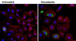
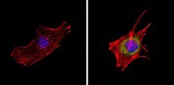
- Immunofluorescent analysis of Cyclin B1 (green) in HeLa cells either left untreated (left panel) or treated with 50nM Nocodazole (right panel) for 16 hours. Formalin fixed cells were permeabilized with 0.1% Triton X-100 in TBS for 10 minutes at room temperature and blocked with 1% Blocker BSA (Product # 37525) for 15 minutes at room temperature. Cells were probed with a Cyclin B1 monoclonal antibody (Product # MA5-14327) at a dilution of 1:100 for at least 1 hour at room temperature, washed with PBS, and incubated with DyLight 488 goat anti-mouse IgG secondary antibody (Product # 35502) at a dilution of 1:400 for 30 minutes at room temperature. F-Actin (red) was stained with DyLight 554 Phalloidin (Product # 21834) and nuclei (blue) were stained with Hoechst 33342 dye (Product # 62249). Images were taken on a Thermo Scientific ArrayScan or ToxInsight Instrument at 20X magnification.

- Submitted by
- Invitrogen Antibodies (provider)
- Main image
- Experimental details
- Immunofluorescent analysis of Cyclin B1 (green) showing staining in the cytoplasm of Hela cells (right) compared to a negative control without primary antibody (left). Formalin-fixed cells were permeabilized with 0.1% Triton X-100 in TBS for 5-10 minutes and blocked with 3% BSA-PBS for 30 minutes at room temperature. Cells were probed with a Cyclin B1 monoclonal antibody (Product # MA5-14327) in 3% BSA-PBS at a dilution of 1:20 and incubated overnight at 4 ºC in a humidified chamber. Cells were washed with PBST and incubated with a DyLight-conjugated secondary antibody in PBS at room temperature in the dark. F-actin (red) was stained with a fluorescent red phalloidin and nuclei (blue) were stained with Hoechst or DAPI. Images were taken at a magnification of 60x.
- Submitted by
- Invitrogen Antibodies (provider)
- Main image

- Experimental details
- Immunofluorescent analysis of Cyclin B1 (green) showing staining in the cytoplasm of HT29 cells (right) compared to a negative control without primary antibody (left). Formalin-fixed cells were permeabilized with 0.1% Triton X-100 in TBS for 5-10 minutes and blocked with 3% BSA-PBS for 30 minutes at room temperature. Cells were probed with a Cyclin B1 monoclonal antibody (Product # MA5-14327) in 3% BSA-PBS at a dilution of 1:20 and incubated overnight at 4 ºC in a humidified chamber. Cells were washed with PBST and incubated with a DyLight-conjugated secondary antibody in PBS at room temperature in the dark. F-actin (red) was stained with a fluorescent red phalloidin and nuclei (blue) were stained with Hoechst or DAPI. Images were taken at a magnification of 60x.
- Submitted by
- Invitrogen Antibodies (provider)
- Main image
- Experimental details
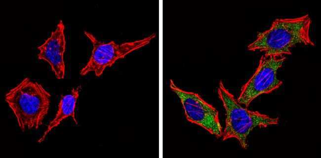
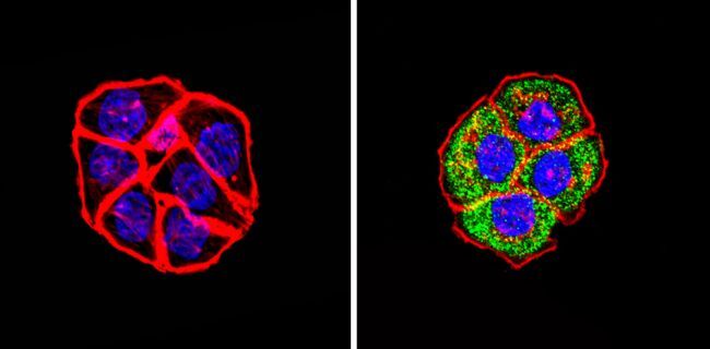
- Immunofluorescent analysis of Cyclin B1 (green) showing staining in the cytoplasm of C2C12 cells (right) compared to a negative control without primary antibody (left). Formalin-fixed cells were permeabilized with 0.1% Triton X-100 in TBS for 5-10 minutes and blocked with 3% BSA-PBS for 30 minutes at room temperature. Cells were probed with a Cyclin B1 monoclonal antibody (Product # MA5-14327) in 3% BSA-PBS at a dilution of 1:20 and incubated overnight at 4 ºC in a humidified chamber. Cells were washed with PBST and incubated with a DyLight-conjugated secondary antibody in PBS at room temperature in the dark. F-actin (red) was stained with a fluorescent red phalloidin and nuclei (blue) were stained with Hoechst or DAPI. Images were taken at a magnification of 60x.

- Submitted by
- Invitrogen Antibodies (provider)
- Main image

- Experimental details
- Immunofluorescent analysis of Cyclin B1 (green) in HeLa cells either left untreated (left panel) or treated with 50nM Nocodazole (right panel) for 16 hours. Formalin fixed cells were permeabilized with 0.1% Triton X-100 in TBS for 10 minutes at room temperature and blocked with 1% Blocker BSA (Product # 37525) for 15 minutes at room temperature. Cells were probed with a Cyclin B1 monoclonal antibody (Product # MA5-14327) at a dilution of 1:100 for at least 1 hour at room temperature, washed with PBS, and incubated with DyLight 488 goat anti-mouse IgG secondary antibody (Product # 35502) at a dilution of 1:400 for 30 minutes at room temperature. F-Actin (red) was stained with DyLight 554 Phalloidin (Product # 21834) and nuclei (blue) were stained with Hoechst 33342 dye (Product # 62249). Images were taken on a Thermo Scientific ArrayScan or ToxInsight Instrument at 20X magnification.

Supportive validation
- Submitted by
- Invitrogen Antibodies (provider)
- Main image
- Experimental details
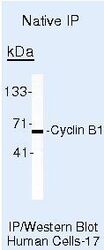
- Immunoprecipitation of Cyclin B1 was performed on the Human Colon Carcinoma cell line LS174T. Antigen-antibody complexes were formed by incubating 500 µL of LS174T whole cell lysate with 5 µL of a Cyclin B1 monoclonal antibody (Product # MA5-14327). The immune complexes were captured using Protein A agarose, washed extensively, and eluted. Samples were resolved on a 4-20% Tris-HCl polyacrylamide gel, transferred to a membrane, and probed with a series of reagents to detect Cyclin B1.
Supportive validation
- Submitted by
- Invitrogen Antibodies (provider)
- Main image
- Experimental details
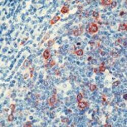
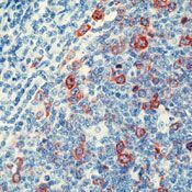
- Immunohistochemistry was performed on formalin-fixed, paraffin-embedded human tonsil tissue. To expose target proteins, heat-induced antigen retrieval was performed by boiling tissue sections in 10mM sodium citrate buffer (pH 6) for 10-20 minutes. Tissues were probed with a Cyclin B1 monoclonal antibody (Product # MA5-14327) at a concentration of 3 µg/mL. Detection was performed using an anti-mouse IgG-HRP secondary antibody followed by a colorimetric substrate.
Supportive validation
- Submitted by
- Invitrogen Antibodies (provider)
- Main image

- Experimental details
- NULL
- Submitted by
- Invitrogen Antibodies (provider)
- Main image
- Experimental details
- NULL
- Submitted by
- Invitrogen Antibodies (provider)
- Main image
- Experimental details
- NULL
- Submitted by
- Invitrogen Antibodies (provider)
- Main image

- Experimental details
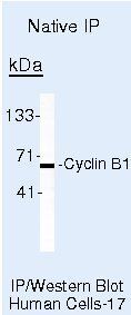
- Immunoprecipitation of Cyclin B1 was performed on the Human Colon Carcinoma cell line LS174T. Antigen-antibody complexes were formed by incubating 500 µL of LS174T whole cell lysate with 5 µL of a Cyclin B1 monoclonal antibody (Product # MA5-14327). The immune complexes were captured using Protein A agarose, washed extensively, and eluted. Samples were resolved on a 4-20% Tris-HCl polyacrylamide gel, transferred to a membrane, and probed with a series of reagents to detect Cyclin B1.
