 Explore
 Validate
 Learn
LearnMA1-10196
antibody from Invitrogen Antibodies
Targeting: EPCAM
17-1A, 323/A3, CD326, CO-17A, EGP-2, EGP34, EGP40, Ep-CAM, ESA, GA733-2, HEA125, KS1/4, KSA, Ly74, M4S1, MH99, MIC18, MK-1, MOC31, TACST-1, TACSTD1, TROP1
 Western blot
 Immunocytochemistry
 Immunoprecipitation
Antibody data
- Antibody Data
- Antigen structure
- References [8]
- Comments [0]
- Validations
- Immunocytochemistry [2]
- Immunohistochemistry [2]
- Flow cytometry [1]
Submit
Validation data
Reference
Comment
Report error
- Product number
- MA1-10196 - Provider product page

- Provider
- Invitrogen Antibodies
- Product name
- EpCAM Monoclonal Antibody (323/A3)
- Antibody type
- Monoclonal
- Antigen
- Other
- Description
- This antibody recognizes an extracellular epitope of CD326 / EpCAM, a marker of epithelial lineages, that is over-expressed in many carcinomas. This antibody will not cross-react with rat. Western Blot: non-reducing conditions
- Reactivity
- Human
- Host
- Mouse
- Isotype
- IgG
- Antibody clone number
- 323/A3
- Vial size
- 100 μg
- Concentration
- 1 mg/mL
- Storage
- 4°C, do not freeze
Submitted references Co-existence of epithelioid and fibroblastoid subsets in a sarcomatoid renal carcinoma cell line revealed by clonal studies.
Enrichment of circulating tumor cells from a large blood volume using leukapheresis and elutriation: proof of concept.
Molecular events associated with epithelial to mesenchymal transition of nasopharyngeal carcinoma cells in the absence of Epstein-Barr virus genome.
Overexpression of epithelial cell adhesion molecule in primary, metastatic, and recurrent/chemotherapy-resistant epithelial ovarian cancer: implications for epithelial cell adhesion molecule-specific immunotherapy.
EpCAM-specific vaccine response by modified antigen and chimeric costimulatory molecule in cynomolgus monkeys.
Gradients in the liver's extracellular matrix chemistry from periportal to pericentral zones: influence on human hepatic progenitors.
Inhibition of proliferation by PERK regulates mammary acinar morphogenesis and tumor formation.
Genetic vaccines against Ep-CAM break tolerance to self in a limited subset of subjects: initial identification of predictive biomarkers.
Hsieh CH, Chen HC, Chang YH, Pang ST, Kuo ML, Chuang CK, Liao SK
Anticancer research 2013 Nov;33(11):4875-89
Anticancer research 2013 Nov;33(11):4875-89
Enrichment of circulating tumor cells from a large blood volume using leukapheresis and elutriation: proof of concept.
Eifler RL, Lind J, Falkenhagen D, Weber V, Fischer MB, Zeillinger R
Cytometry. Part B, Clinical cytometry 2011 Mar;80(2):100-11
Cytometry. Part B, Clinical cytometry 2011 Mar;80(2):100-11
Molecular events associated with epithelial to mesenchymal transition of nasopharyngeal carcinoma cells in the absence of Epstein-Barr virus genome.
Lin JC, Liao SK, Lee EH, Hung MS, Sayion Y, Chen HC, Kang CC, Huang LS, Cherng JM
Journal of biomedical science 2009 Nov 24;16:105
Journal of biomedical science 2009 Nov 24;16:105
Overexpression of epithelial cell adhesion molecule in primary, metastatic, and recurrent/chemotherapy-resistant epithelial ovarian cancer: implications for epithelial cell adhesion molecule-specific immunotherapy.
Bellone S, Siegel ER, Cocco E, Cargnelutti M, Silasi DA, Azodi M, Schwartz PE, Rutherford TJ, Pecorelli S, Santin AD
International journal of gynecological cancer : official journal of the International Gynecological Cancer Society 2009 Jul;19(5):860-6
International journal of gynecological cancer : official journal of the International Gynecological Cancer Society 2009 Jul;19(5):860-6
EpCAM-specific vaccine response by modified antigen and chimeric costimulatory molecule in cynomolgus monkeys.
Neighbors M, Apt D, Chang JC, Brinkman A, Sipos-Solman I, Ong R, Leong S, Punnonen J
Journal of immunotherapy (Hagerstown, Md. : 1997) 2008 Sep;31(7):644-55
Journal of immunotherapy (Hagerstown, Md. : 1997) 2008 Sep;31(7):644-55
Gradients in the liver's extracellular matrix chemistry from periportal to pericentral zones: influence on human hepatic progenitors.
McClelland R, Wauthier E, Uronis J, Reid L
Tissue engineering. Part A 2008 Jan;14(1):59-70
Tissue engineering. Part A 2008 Jan;14(1):59-70
Inhibition of proliferation by PERK regulates mammary acinar morphogenesis and tumor formation.
Sequeira SJ, Ranganathan AC, Adam AP, Iglesias BV, Farias EF, Aguirre-Ghiso JA
PloS one 2007 Jul 18;2(7):e615
PloS one 2007 Jul 18;2(7):e615
Genetic vaccines against Ep-CAM break tolerance to self in a limited subset of subjects: initial identification of predictive biomarkers.
Elia L, Mennuni C, Storto M, Podda S, Calvaruso F, Salucci V, Aurisicchio L, Scarito A, Ciliberto G, La Monica N, Palombo F
European journal of immunology 2006 May;36(5):1337-49
European journal of immunology 2006 May;36(5):1337-49
No comments: Submit comment
Supportive validation
- Submitted by
- Invitrogen Antibodies (provider)
- Main image
- Experimental details
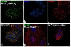
- Immunofluorescence analysis of EpCAM was performed using HT-29 and HeLa cells. The cells were fixed with 4% paraformaldehyde for 10 minutes, permeabilized with 0.1% Triton™ X-100 for 15 minutes, and blocked with 2% BSA for 1 hour at room temperature. The cells were labeled with EpCAM Mouse Monoclonal Antibody (Product # MA1-10196) at 1 µg/mL in 0.1% BSA and incubated overnight at 4 degree and then labeled with Goat anti-Mouse IgG (H+L) Highly Cross-Adsorbed Secondary Antibody, Alexa Fluor Plus 488 (Product # A32723) at a dilution of 1:2000 for 45 minutes at room temperature (Panel a: green) in HT-29 cells. Nuclei (Panel b: blue) were stained with ProLong™ Diamond Antifade Mountant with DAPI (Product # P36962). F-actin (Panel c: red) was stained with Rhodamine Phalloidin (Product # R415, 1:300). Panel d represents the merged image of HT-29 cells, which is a positive model for EpCAM expression showing a plasma membrane localization. Panel e represents the merged image of HeLa cells, that are null for EpCAM protein expression. Panel f represents control cells with no primary antibody to assess background. The images were captured at 60X magnification.
- Submitted by
- Invitrogen Antibodies (provider)
- Main image

- Experimental details
- Immunofluorescence analysis of EpCAM was performed using HT-29 and HeLa cells. The cells were fixed with 4% paraformaldehyde for 10 minutes, permeabilized with 0.1% Triton™ X-100 for 15 minutes, and blocked with 2% BSA for 1 hour at room temperature. The cells were labeled with EpCAM Mouse Monoclonal Antibody (Product # MA1-10196) at 1 µg/mL in 0.1% BSA and incubated overnight at 4 degree and then labeled with Goat anti-Mouse IgG (H+L) Highly Cross-Adsorbed Secondary Antibody, Alexa Fluor Plus 488 (Product # A32723) at a dilution of 1:2000 for 45 minutes at room temperature (Panel a: green) in HT-29 cells. Nuclei (Panel b: blue) were stained with ProLong™ Diamond Antifade Mountant with DAPI (Product # P36962). F-actin (Panel c: red) was stained with Rhodamine Phalloidin (Product # R415, 1:300). Panel d represents the merged image of HT-29 cells, which is a positive model for EpCAM expression showing a plasma membrane localization. Panel e represents the merged image of HeLa cells, that are null for EpCAM protein expression. Panel f represents control cells with no primary antibody to assess background. The images were captured at 60X magnification.

Supportive validation
- Submitted by
- Invitrogen Antibodies (provider)
- Main image
- Experimental details
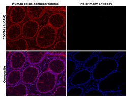
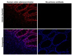
- Immunohistochemical analysis of CD326 (EpCAM) was performed using formalin-fixed paraffin-embedded human colon adenocarcinoma tissue sections. To expose the target protein, heat-induced epitope retrieval was performed on de-paraffinized sections using eBioscience™ IHC Antigen Retrieval Solution - Low pH (10X) (Product # 00-4955-58) diluted to 1X solution in water in a decloaking chamber at 110 degree Celsius for 15 minutes. Following antigen retrieval, the sections were blocked with 3% H2O2 for 1 h at room temperature followed by 2% normal goat serum in 1X PBS for 45 minutes at room temperature. The sections were then probed with or without EpCAM Monoclonal Antibody (323/A3) (Product # MA1-10196) at a concentration of 2 µg/mL in 0.1% normal goat serum overnight at 4 degree Celsius in a humidified chamber. Detection was performed using Alexa Fluor™ 647 Tyramide SuperBoost™ Kit, goat anti-mouse IgG (Product # B40916). Nuclei were stained with DAPI (Product # D1306) and the sections were mounted using ProLong™ Glass Antifade Mountant (Product # P36984). The images were captured on EVOS™ M7000 Imaging System (Product # AMF7000) at 20X magnification and externally deconvoluted.
- Submitted by
- Invitrogen Antibodies (provider)
- Main image
- Experimental details
- Immunohistochemical analysis of CD326 (EpCAM) was performed using formalin-fixed paraffin-embedded human colon adenocarcinoma tissue sections. To expose the target protein, heat-induced epitope retrieval was performed on de-paraffinized sections using eBioscience™ IHC Antigen Retrieval Solution - Low pH (10X) (Product # 00-4955-58) diluted to 1X solution in water in a decloaking chamber at 110 degree Celsius for 15 minutes. Following antigen retrieval, the sections were blocked with 3% H2O2 for 1 h at toom temperature followed by 2% normal goat serum in 1X PBS for 45 minutes at room temperature. The sections were then probed with or without EpCAM Monoclonal Antibody (323/A3) (Product # MA1-10196) at a concentration of 2 µg/mL in 0.1% normal goat serum overnight at 4 degree Celsius in a humidified chamber. Detection was performed using Alexa Fluor™ 647 Tyramide SuperBoost™ Kit, goat anti-mouse IgG (Product # B40916). Nuclei were stained with DAPI (Product # D1306) and the sections were mounted using ProLong™ Glass Antifade Mountant (Product # P36984). The images were captured on EVOS™ M7000 Imaging System (Product # AMF7000) at 20X magnification and externally deconvoluted.

Supportive validation
- Submitted by
- Invitrogen Antibodies (provider)
- Main image
- Experimental details
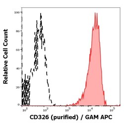
- Flow Cytometry analysis of EpCAM using EpCAM Monoclonal Antibody (323/A3) (Product # MA1-10196). Separation of MCF-7 cells (red-filled) from Jurkat cells (black-dashed) in flow cytometry analysis (surface staining) of cellular suspensions of Jurkat and MCF-7 cell lines stained using EpCAM (323/A3) purified antibody (concentration in sample 1 μg/mL, GAM APC).
