 Explore
 Validate
 Learn
LearnMAB960-100
antibody from R&D Systems
Targeting: EPCAM
17-1A, 323/A3, CD326, CO-17A, EGP-2, EGP34, EGP40, Ep-CAM, ESA, GA733-2, HEA125, KS1/4, KSA, Ly74, M4S1, MH99, MIC18, MK-1, MOC31, TACST-1, TACSTD1, TROP1
 Western blot
 Immunocytochemistry
Antibody data
- Antibody Data
- Antigen structure
- References [6]
- Comments [0]
- Validations
- Immunocytochemistry [2]
- Immunohistochemistry [1]
Submit
Validation data
Reference
Comment
Report error
- Product number
- MAB960-100 - Provider product page

- Provider
- R&D Systems
- Product name
- Human EpCAM/TROP-1 Antibody
- Antibody type
- Monoclonal
- Description
- Protein A or G purified from hybridoma culture supernatant. Detects human EpCAM in direct ELISAs and Western blots. This antibody detects an epitope found in the extracellular domain between amino acids 136 and 265. In direct ELISAs and Western blots, no cross-reactivity with recombinant human (rh) ALCAM, rhBCAM, rhMCAM, rhNCAM-L1, or rmOCAM is observed.
- Reactivity
- Human
- Host
- Mouse
- Conjugate
- Unconjugated
- Antigen sequence
P16422- Isotype
- IgG
- Antibody clone number
- 158210
- Vial size
- 100 ug
- Storage
- Use a manual defrost freezer and avoid repeated freeze-thaw cycles. 12 months from date of receipt, -20 to -70 °C as supplied. 1 month, 2 to 8 °C under sterile conditions after reconstitution. 6 months, -20 to -70 °C under sterile conditions after reconstitution.
Submitted references Polymeric mechanical amplifiers of immune cytokine-mediated apoptosis.
EpCAM-Regulated Transcription Exerts Influences on Nanomechanical Properties of Endometrial Cancer Cells That Promote Epithelial-to-Mesenchymal Transition.
Identification of tumorigenic cells and therapeutic targets in pancreatic neuroendocrine tumors.
Integrative genomics identifies YY1AP1 as an oncogenic driver in EpCAM(+) AFP(+) hepatocellular carcinoma.
Detection of brain tumor cells in the peripheral blood by a telomerase promoter-based assay.
Activation of hepatic stem cell marker EpCAM by Wnt-beta-catenin signaling in hepatocellular carcinoma.
Mitchell MJ, Webster J, Chung A, Guimarães PP, Khan OF, Langer R
Nature communications 2017 Mar 20;8:14179
Nature communications 2017 Mar 20;8:14179
EpCAM-Regulated Transcription Exerts Influences on Nanomechanical Properties of Endometrial Cancer Cells That Promote Epithelial-to-Mesenchymal Transition.
Hsu YT, Osmulski P, Wang Y, Huang YW, Liu L, Ruan J, Jin VX, Kirma NB, Gaczynska ME, Huang TH
Cancer research 2016 Nov 1;76(21):6171-6182
Cancer research 2016 Nov 1;76(21):6171-6182
Identification of tumorigenic cells and therapeutic targets in pancreatic neuroendocrine tumors.
Krampitz GW, George BM, Willingham SB, Volkmer JP, Weiskopf K, Jahchan N, Newman AM, Sahoo D, Zemek AJ, Yanovsky RL, Nguyen JK, Schnorr PJ, Mazur PK, Sage J, Longacre TA, Visser BC, Poultsides GA, Norton JA, Weissman IL
Proceedings of the National Academy of Sciences of the United States of America 2016 Apr 19;113(16):4464-9
Proceedings of the National Academy of Sciences of the United States of America 2016 Apr 19;113(16):4464-9
Integrative genomics identifies YY1AP1 as an oncogenic driver in EpCAM(+) AFP(+) hepatocellular carcinoma.
Zhao X, Parpart S, Takai A, Roessler S, Budhu A, Yu Z, Blank M, Zhang YE, Jia HL, Ye QH, Qin LX, Tang ZY, Thorgeirsson SS, Wang XW
Oncogene 2015 Sep 24;34(39):5095-104
Oncogene 2015 Sep 24;34(39):5095-104
Detection of brain tumor cells in the peripheral blood by a telomerase promoter-based assay.
Macarthur KM, Kao GD, Chandrasekaran S, Alonso-Basanta M, Chapman C, Lustig RA, Wileyto EP, Hahn SM, Dorsey JF
Cancer research 2014 Apr 15;74(8):2152-9
Cancer research 2014 Apr 15;74(8):2152-9
Activation of hepatic stem cell marker EpCAM by Wnt-beta-catenin signaling in hepatocellular carcinoma.
Yamashita T, Budhu A, Forgues M, Wang XW
Cancer research 2007 Nov 15;67(22):10831-9
Cancer research 2007 Nov 15;67(22):10831-9
No comments: Submit comment
Supportive validation
- Submitted by
- R&D Systems (provider)
- Main image
- Experimental details
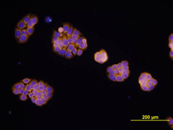
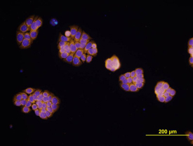
- EpCAM/TROP-1 in HT-29 Human Cell Line. EpCAM/TROP-1 was detected in immersion fixed HT-29 human colon adenocarcinoma cell line using Mouse Anti-Human EpCAM/TROP-1 Monoclonal Antibody (Catalog # MAB960) at 10 µg/mL for 3 hours at room temperature. Cells were stained using the NorthernLights™ 557-conjugated Anti-Mouse IgG Secondary Antibody (yellow; Catalog # NL007) and counterstained with DAPI (blue). View our protocol for Fluorescent ICC Staining of Cells on Coverslips.
- Submitted by
- R&D Systems (provider)
- Main image
- Experimental details
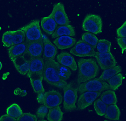
- EpCAM/TROP-1 in HT-29 Human Cell Line. EpCAM/TROP-1 was detected in immersion fixed HT-29 human colon adenocarcinoma cell line using Mouse Anti-Human EpCAM/TROP-1 Monoclonal Antibody (Catalog # MAB960) at 10 µg/mL for 3 hours at room temperature. Cells were stained using the NorthernLights™ 493-conjugated Anti-Mouse IgG Secondary Antibody (green; Catalog # NL009) and counterstained with DAPI (blue). View our protocol for Fluorescent ICC Staining of Cells on Coverslips.

Supportive validation
- Submitted by
- R&D Systems (provider)
- Main image
- Experimental details
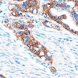
- EpCAM/TROP-1 in Human Adenocarcinoma. EpCAM/TROP-1 was detected in immersion fixed paraffin-embedded sections of human adenocarcinoma using Mouse Anti-Human EpCAM/TROP-1 Monoclonal Antibody (Catalog # MAB960) at 15 µg/mL overnight at 4 °C. Tissue was stained using the Anti-Mouse HRP-DAB Cell & Tissue Staining Kit (brown; Catalog # CTS002) and counterstained with hematoxylin (blue). Specific staining was localized to cancer cells. View our protocol for Chromogenic IHC Staining of Paraffin-embedded Tissue Sections.
