Explore
 Validate
 Learn
LearnMA5-29246
antibody from Invitrogen Antibodies
Targeting: EPCAM
17-1A, 323/A3, CD326, CO-17A, EGP-2, EGP34, EGP40, Ep-CAM, ESA, GA733-2, HEA125, KS1/4, KSA, Ly74, M4S1, MH99, MIC18, MK-1, MOC31, TACST-1, TACSTD1, TROP1
 Western blot
 ELISA
ELISA Immunocytochemistry Immunoprecipitation Immunohistochemistry Flow cytometry Other assay
Antibody data
- Antibody Data
- Antigen structure
- References [1]
- Comments [0]
- Validations
- Immunocytochemistry [3]
- Immunoprecipitation [1]
- Immunohistochemistry [2]
- Flow cytometry [3]
- Other assay [2]
Submit
Validation data
Reference
Comment
Report error
- Product number
- MA5-29246 - Provider product page

- Provider
- Invitrogen Antibodies
- Product name
- EpCAM Recombinant Rabbit Monoclonal Antibody (28)
- Antibody type
- Monoclonal
- Antigen
- Recombinant full-length protein
- Description
- This product is preservative free. It is recommended to add sodium azide to avoid contamination (final concentration 0.05%-0.1%). Recombinant rabbit monoclonal antibodies are produced using in vitro expression systems. The expression systems are developed by cloning in the specific antibody DNA sequences from immunoreactive rabbits. Then, individual clones are screened to select the best candidates for production. The advantages of using recombinant rabbit monoclonal antibodies include: better specificity and sensitivity, lot-to-lot consistency, animal origin-free formulations, and broader immunoreactivity to diverse targets due to larger rabbit immune repertoire. This antibody has specificity for Human EpCAM/TROP1/CD326.
- Reactivity
- Human
- Host
- Rabbit
- Isotype
- IgG
- Antibody clone number
- 28
- Vial size
- 100 μL
- Concentration
- 1.12 mg/mL
- Storage
- Store at 4°C short term. For long term storage, store at -20°C, avoiding freeze/thaw cycles.
Submitted references High throughput, label-free isolation of circulating tumor cell clusters in meshed microwells.
Boya M, Ozkaya-Ahmadov T, Swain BE, Chu CH, Asmare N, Civelekoglu O, Liu R, Lee D, Tobia S, Biliya S, McDonald LD, Nazha B, Kucuk O, Sanda MG, Benigno BB, Moreno CS, Bilen MA, McDonald JF, Sarioglu AF
Nature communications 2022 Jun 13;13(1):3385
Nature communications 2022 Jun 13;13(1):3385
No comments: Submit comment
Supportive validation
- Submitted by
- Invitrogen Antibodies (provider)
- Main image

- Experimental details
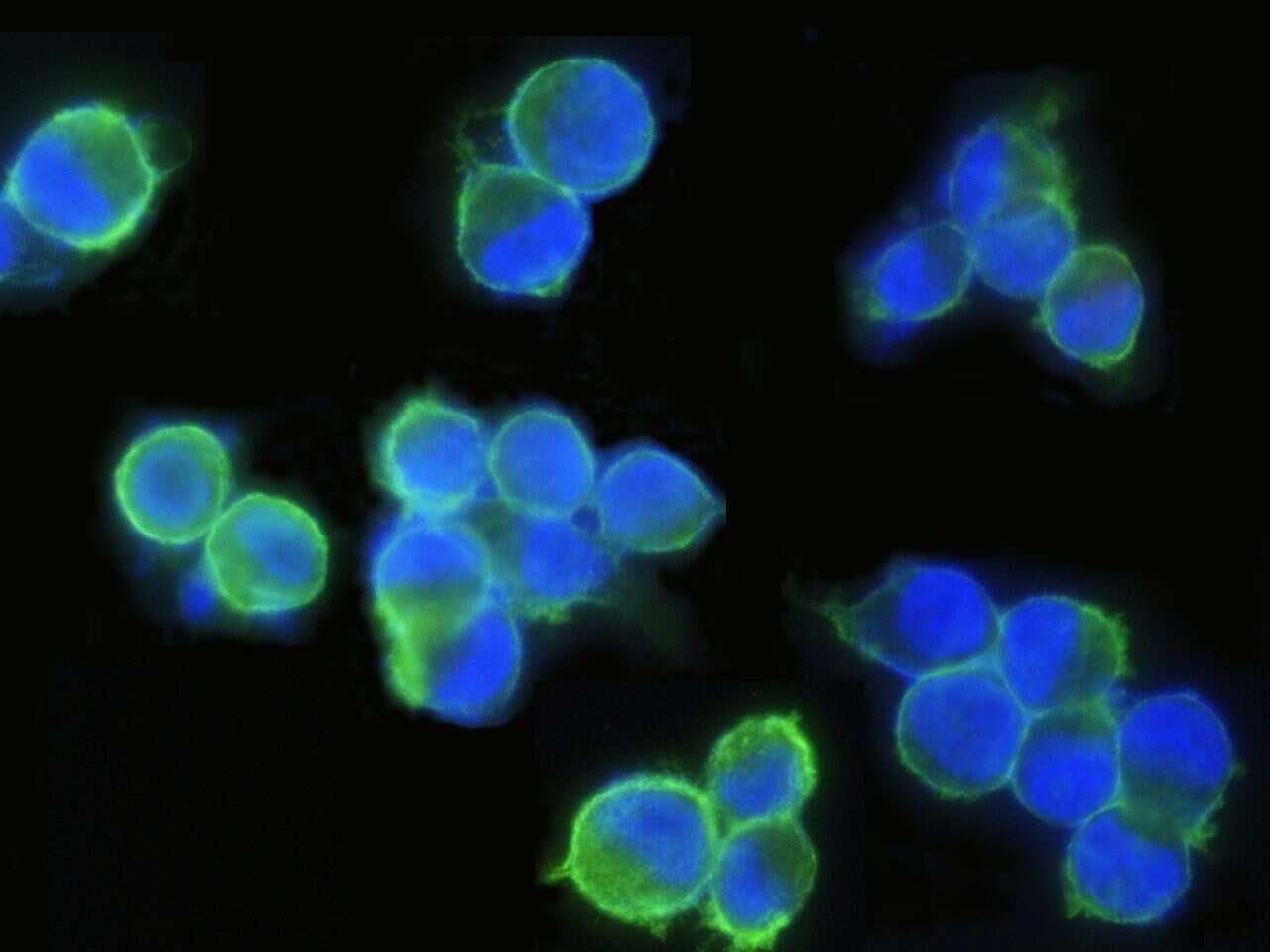
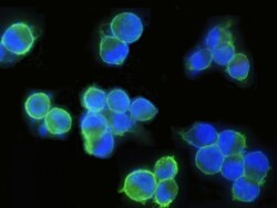
- Immunofluorescence staining of Human EpCAM in SKBR3 cells. Cells were fixed with 4% PFA, blocked with 10% serum, and incubated with EpCAM Recombinant Rabbit Monoclonal Antibody (28) (Product # MA5-29246, 1:500) at 4°C overnight. Then cells were stained with the Alexa Fluor® 488-conjugated Goat Anti-rabbit IgG secondary antibody (green) and counterstained with DAPI (blue). Positive staining was localized to plasma membrane.
- Submitted by
- Invitrogen Antibodies (provider)
- Main image
- Experimental details
- Immunofluorescence staining of Human EpCAM in SKBR3 cells. Cells were fixed with 4% PFA, blocked with 10% serum, and incubated with EpCAM Recombinant Rabbit Monoclonal Antibody (28) (Product # MA5-29246, 1:500) at 4°C overnight. Then cells were stained with the Alexa Fluor® 488-conjugated Goat Anti-rabbit IgG secondary antibody (green) and counterstained with DAPI (blue). Positive staining was localized to plasma membrane.

- Submitted by
- Invitrogen Antibodies (provider)
- Main image

- Experimental details
- Immunofluorescence staining of Human EpCAM in SKBR3 cells. Cells were fixed with 4% PFA, blocked with 10% serum, and incubated with EpCAM Recombinant Rabbit Monoclonal Antibody (28) (Product # MA5-29246, 1:500) at 4°C overnight. Then cells were stained with the Alexa Fluor® 488-conjugated Goat Anti-rabbit IgG secondary antibody (green) and counterstained with DAPI (blue). Positive staining was localized to plasma membrane.

Supportive validation
- Submitted by
- Invitrogen Antibodies (provider)
- Main image

- Experimental details
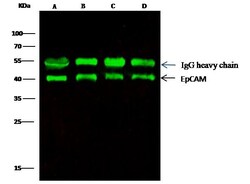
- EpCAM Immunoprecipitation using: Lane A: 0.5 mg MCF-7 Whole Cell Lysate, Lane B: 0.5 mg A431 Whole Cell Lysate, Lane C: 0.5 mg HepG2 Whole Cell Lysate, Lane D: 0.5 mg Caco-2 Whole Cell Lysate 0.5 µL with EpCAM Recombinant Rabbit Monoclonal Antibody (28) (Product # MA5-29246) and 15 µL of 50 % Protein G agarose. Primary antibody: EpCAM Recombinant Rabbit Monoclonal Antibody (28), at 1:5,000 dilution. Secondary antibody: Dylight 800-labeled antibody to rabbit IgG (H+L), at 1:5,000 dilution. Developed using the Odyssey technique. Performed under reducing conditions. Predicted band size: 40 kDa. Observed band size: 40 kDa.

Supportive validation
- Submitted by
- Invitrogen Antibodies (provider)
- Main image

- Experimental details
- Immunohistochemical staining of human EpCAM in human colon carcinoma with EpCAM Recombinant Rabbit Monoclonal Antibody (28) (Product # MA5-29246, 1:2,500 dilution, formalin-fixed paraffin embedded sections). Positive staining was localized to membrane of colonic gland epithelium.

- Submitted by
- Invitrogen Antibodies (provider)
- Main image

- Experimental details
- Immunohistochemical staining of human EpCAM in human bladder carcinoma with EpCAM Recombinant Rabbit Monoclonal Antibody (28) (Product # MA5-29246, 1:2,500 dilution, formalin-fixed paraffin embedded sections). Positive staining was localized to membrane of transitional epithelium.

Supportive validation
- Submitted by
- Invitrogen Antibodies (provider)
- Main image
- Experimental details
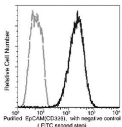
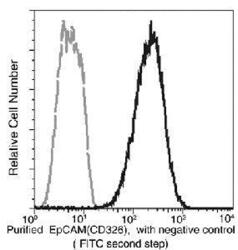
- Flow cytometric analysis of anti-EpCAM (CD326) reactivity on SKBR3 cells. SKBR3 cells were stained with EpCAM Monoclonal Antibody (Product # MA5-29246), then a FITC-conjugated second step antibody. The histogram were derived from the gated events based on light scattering characteristics of viable cells.

- Submitted by
- Invitrogen Antibodies (provider)
- Main image
- Experimental details
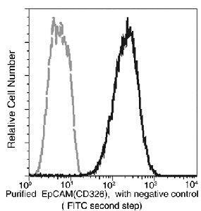
- Flow cytometric analysis of EpCAM (CD326) in SKBR3 cells. SKBR3 cells were stained with EpCAM Recombinant Rabbit Monoclonal Antibody (28) (Product # MA5-29246), then a FITC-conjugated Secondary antibody. The histogram were derived from the gated events based on light scattering characteristics of viable cells.
- Submitted by
- Invitrogen Antibodies (provider)
- Main image

- Experimental details
- Flow cytometric analysis of EpCAM (CD326) in SKBR3 cells. SKBR3 cells were stained with EpCAM Recombinant Rabbit Monoclonal Antibody (28) (Product # MA5-29246), then a FITC-conjugated Secondary antibody. The histogram were derived from the gated events based on light scattering characteristics of viable cells.

Supportive validation
- Submitted by
- Invitrogen Antibodies (provider)
- Main image
- Experimental details
- EpCAM Immunoprecipitation using: Lane A: 0.5 mg MCF-7 Whole Cell Lysate, Lane B: 0.5 mg A431 Whole Cell Lysate, Lane C: 0.5 mg HepG2 Whole Cell Lysate, Lane D: 0.5 mg Caco-2 Whole Cell Lysate 0.5 µL with EpCAM Recombinant Rabbit Monoclonal Antibody (28) (Product # MA5-29246) and 15 µL of 50 % Protein G agarose. Primary antibody: EpCAM Recombinant Rabbit Monoclonal Antibody (28), at 1:5,000 dilution. Secondary antibody: Dylight 800-labeled antibody to rabbit IgG (H+L), at 1:5,000 dilution. Developed using the Odyssey technique. Performed under reducing conditions. Predicted band size: 40 kDa. Observed band size: 40 kDa.

- Submitted by
- Invitrogen Antibodies (provider)
- Main image
- Experimental details
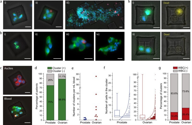
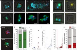
- Isolation of CTC clusters from ovarian and prostate cancer patients. a Fluorescence microscope images of the isolated CTC clusters from ovarian cancer patients. Captured cells were stained with Cytokeratin, Vimentin (green), CD45 (red), and DAPI (nuclei, blue). Scale bars, 20 mum. b Images of the isolated CTC clusters from metastatic prostate cancer patients. Captured cells were stained with Cytokeratin, Vimentin, PSA/KLK3, EpCAM (green), CD45(red), and DAPI (nuclei, blue). Scale bars, 20 mum. c Clusters isolated from the peripheral blood and ascites sample drawn from the peritoneal cavity of an ovarian cancer patient at the same timepoint. The isolated CTC cluster was morphologically different compared to the tumor spheroids isolated from ascites sample of the same patient. The spheroids from ascites sample were fixed and stained with Cytokeratin, EpCAM (red), CD45 (green) and DAPI (nuclei, blue), and CTC clusters isolated from blood sample were fixed and stained with Cytokeratin, Vimentin (green), CD45 (red) and DAPI (nuclei, blue). Scale bars, 20 mum. d Percentage of prostate ( n = 8) and ovarian ( n = 9) cancer patients that are observed to have CTC cluster(s) in their blood samples. e Number of CTC clusters observed per milliliter of blood samples from prostate and ovarian cancer patients. f Distribution of the number of cells observed in the prostate and ovarian CTC clusters. The box plots show the 25th and 75th percentiles, line shows median, and whiskers show maxima a