 Explore
 Validate
 Learn
LearnMA5-15640
antibody from Invitrogen Antibodies
Targeting: EPCAM
17-1A, 323/A3, CD326, CO-17A, EGP-2, EGP34, EGP40, Ep-CAM, ESA, GA733-2, HEA125, KS1/4, KSA, Ly74, M4S1, MH99, MIC18, MK-1, MOC31, TACST-1, TACSTD1, TROP1
 Western blot
Antibody data
- Antibody Data
- Antigen structure
- References [2]
- Comments [0]
- Validations
- Western blot [5]
- Immunocytochemistry [1]
- Immunohistochemistry [1]
Submit
Validation data
Reference
Comment
Report error
- Product number
- MA5-15640 - Provider product page

- Provider
- Invitrogen Antibodies
- Product name
- EpCAM Monoclonal Antibody (7E11)
- Antibody type
- Monoclonal
- Antigen
- Purifed from natural sources
- Description
- MA5-15640 targets EPCAM in IHC and WB applications and shows reactivity with Human samples.
- Antibody clone number
- 7E11
- Concentration
- Conc. Not Determined
Submitted references Epstein-Barr virus-associated lymphoepithelioma-like cholangiocarcinoma: a rare variant of intrahepatic cholangiocarcinoma with favourable outcome.
Expression of stemness markers (CD133 and EpCAM) in prognostication of hepatocellular carcinoma.
Chan AW, Tong JH, Sung MY, Lai PB, To KF
Histopathology 2014 Nov;65(5):674-83
Histopathology 2014 Nov;65(5):674-83
Expression of stemness markers (CD133 and EpCAM) in prognostication of hepatocellular carcinoma.
Chan AW, Tong JH, Chan SL, Lai PB, To KF
Histopathology 2014 Jun;64(7):935-50
Histopathology 2014 Jun;64(7):935-50
No comments: Submit comment
Supportive validation
- Submitted by
- Invitrogen Antibodies (provider)
- Main image

- Experimental details
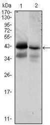
- Western blot analysis of CD326/EpCAM/Epithelial Specific antigen using CD326/EpCAM/Epithelial Specific antigen monoclonal antibody (Product # MA5-15640) in HTC116 (1) and T47D (2) cell lysate.

- Submitted by
- Invitrogen Antibodies (provider)
- Main image
- Experimental details
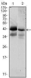
- Western blot analysis of CD326/EpCAM/Epithelial Specific antigen using CD326/EpCAM/Epithelial Specific antigen monoclonal antibody (Product # MA5-15640) in HTC116 (1) and T47D (2) cell lysate.
- Submitted by
- Invitrogen Antibodies (provider)
- Main image

- Experimental details
- Western blot analysis of CD326/EpCAM/Epithelial Specific antigen using CD326/EpCAM/Epithelial Specific antigen monoclonal antibody (Product # MA5-15640) in HTC116 (1) and T47D (2) cell lysate.

- Submitted by
- Invitrogen Antibodies (provider)
- Main image

- Experimental details
- Knockout of EpCAM was achieved by CRISPR-Cas9 genome editing using LentiArray™ Lentiviral sgRNA (Product # A32042, Assay ID CRISPR701274_LV) and LentiArray Cas9 Lentivirus (Product # A32064). Western blot analysis of EpCAM was performed by loading 30 µg of A-431 wild type (Lane 1), A-431 Cas9 (Lane 2) andA-431 EpCAM KO (Lane 3) membrane enriched extracts. The samples were electrophoresed using NuPAGE™ Novex™ 4-12% Bis-Tris Protein Gel (Product # NP0322BOX). Resolved proteins were then transferred onto a nitrocellulose membrane (Product # IB23001) by iBlot® 2 Dry Blotting System (Product # IB21001). The blot was probed with Anti-EpCAM Monoclonal Antibody (7E11) (Product # MA5-15640, 1:1000 dilution) and Goat anti-Mouse IgG (H+L) Superclonal™ Recombinant Secondary Antibody, HRP (Product # A28177, 1:5000 dilution) using the iBright FL 1000 (Product # A32752). Chemiluminescent detection was performed using SuperSignal™ West Dura Extended Duration Substrate (Product # 34076). Loss of signal upon CRISPR mediated knockout (KO) using the LentiArray™ CRISPR product line confirms that antibody is specific to EpCAM.

- Submitted by
- Invitrogen Antibodies (provider)
- Main image
- Experimental details
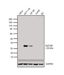
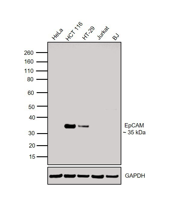
- Western blot was performed using Anti-EpCAM Mouse Monoclonal Antibody (Product # MA5-15640) and a 35 kDa band corresponding to EpCAM was observed in cell lines tested except for HeLa, Jurkat and BJ. Membrane enriched extracts (30 µg lysate) of HeLa (Lane 1), HCT 116 (Lane 2), HT-29 (Lane 3), Jurkat (Lane 4) and BJ (Lane 5) were electrophoresed using Novex® NuPAGE® 4-12% Bis-Tris Protein Gel (Product # NP0322BOX). Resolved proteins were then transferred onto a nitrocellulose membrane (Product # IB23001) by iBlot® 2 Dry Blotting System (Product # IB21001). The blot was probed with the primary antibody (1:1000 dilution) and detected by detected by chemiluminescence with Goat anti-Mouse IgG (H+L), Superclonal™ Recombinant Secondary Antibody, HRP (Product # A28177, 1:4000 dilution) using the iBright FL 1000 (Product # A32752). Chemiluminescent detection was performed using Novex® ECL Chemiluminescent Substrate Reagent Kit (Product # WP20005).
Supportive validation
- Submitted by
- Invitrogen Antibodies (provider)
- Main image

- Experimental details
- Immunofluorescence analysis of EpCAM was performed using HT-29 and HeLa cells. The cells were fixed with 4% paraformaldehyde for 10 minutes, permeabilized with 0.1% Triton™ X-100 for 15 minutes, and blocked with 2% BSA for 1 hour at room temperature. The cells were labeled with EpCAM Mouse Monoclonal Antibody (Product # MA5-15640) at 1:100 dilution in 0.1% BSA and incubated overnight at 4 degree and then labeled with Goat anti-Mouse IgG (H+L) Highly Cross-Adsorbed Secondary Antibody, Alexa Fluor Plus 488 (Product # A32723) at a dilution of 1:2000 for 45 minutes at room temperature (Panel a: green) in HT-29 cells. Nuclei (Panel b: blue) were stained with ProLong™ Diamond Antifade Mountant with DAPI (Product # P36962). F-actin (Panel c: red) was stained with Rhodamine Phalloidin (Product # R415, 1:300). Panel d represents the merged image of HT-29 cells, which is a positive model for EpCAM expression showing a plasma membrane localization. Panel e represents the merged image of HeLa cells, that are null for EpCAM protein expression. Panel f represents control cells with no primary antibody to assess background. The images were captured at 60X magnification.

Supportive validation
- Submitted by
- Invitrogen Antibodies (provider)
- Main image
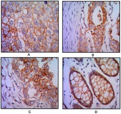
- Experimental details
- Immunohistochemical analysis of paraffin-embedded human lung cancer (A), colon cancer (B), breast cancer (C) and rectal cancer (D), using CD326/EpCAM/Epithelial Specific antigen monoclonal antibody (Product # MA5-15640) followed with DAB staining.
