Explore
 Validate
 Learn
Learn Western blot
Antibody data
- Antibody Data
- Antigen structure
- References [0]
- Comments [0]
- Validations
- Western blot [2]
- Immunocytochemistry [2]
- Flow cytometry [1]
Submit
Validation data
Reference
Comment
Report error
- Product number
- 44-454G - Provider product page

- Provider
- Invitrogen Antibodies
- Product name
- Phospho-MEK1/MEK2 (Ser218, Ser222, Ser226) Polyclonal Antibody
- Antibody type
- Polyclonal
- Antigen
- Synthetic peptide
- Reactivity
- Human, Mouse, Rat
- Host
- Rabbit
- Isotype
- IgG
- Vial size
- 100 µL
- Storage
- -20°C
No comments: Submit comment
Supportive validation
- Submitted by
- Invitrogen Antibodies (provider)
- Main image
- Experimental details
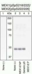
- Peptide Competition and Stimulation Extracts of HeLa cells untreated (1) or treated with 200 ng/mL PMA for 10 minutes (2-5) were resolved by SDS-PAGE on a 10% Tris-glycine gel and transferred to PVDF. The membrane was blocked with a 5% milk-TBST buffer for 1 hour at RT, then incubated with the MEK1 (pSpS218/222)/MEK2 (pSpS222/226) antibody at 4°C in a 3% milk-TBST buffer, following prior incubation with: no peptide (1, 2), the non-phosphopeptide corresponding to the phosphopeptide immunogen (3), a generic phosphoserine-containing peptide (4), or the phosphopeptide immunogen (5). After washing, the membrane was incubated with goat F (ab’)2 anti-rabbit IgG alkaline phosphatase (Product # ALI4405) and signals were detected using the Pierce SuperSignal™ method. The data show that only the phosphopeptide corresponding to MEK1 (pSpS218/222)/MEK2 (pSpS222/226) blocks the antibody signal, demonstrating the specificity of the antibody. The data also show the induction of MEK1 (pSpS218/222)/MEK2 (pSpS222/226) phosphorylation by the addition of PMA to this cell system.

- Submitted by
- Invitrogen Antibodies (provider)
- Main image
- Experimental details
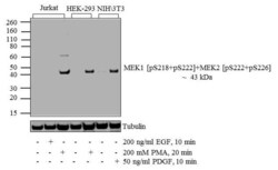
- Western blot analysis of MEK1 (pS218)/ (pS222) + MEK2 (pS222)/ (pS226) was performed by loading 20 µg of Jurkat (lane1), Jurkat treated for 10 minutes with 200 ng/mL of EGF (lane2), Jurkat treated for 20 minutes with 200 nM of PMA (lane3), HEK-293 (lane4), HEK-293 treated for 20 minutes with 200 nM of PMA (lane5), NIH\3T3 (lane6) and NIH\3T3 treated for 10 minutes with 50 ng/mL of PDGF (lane7) cell lysate using Novex® NuPAGE® 4-12 % Bis-Tris gel (Product # NP0322BOX), XCell SureLock™ Electrophoresis System (Product # EI0002), Novex® Sharp Pre-Stained Protein Standard (LC5800), and iBlot® 2 Dry Blotting System (IB21001). Proteins were transferred to a nitrocellulose membrane and blocked with 5 % skim milk for 1 hour at room temperature. MEK1 (pS218)/ (pS222) + MEK2 (pS222)/ (pS226) was detected at ~ 43 kDa using MEK1 (pS218)/ (pS222) + MEK2 (pS222)/ (pS226) Rabbit Polyclonal Antibody (Product # 44-244G) at 1:1000 dilution in 5 % skim milk at 4°C overnight on a rocking platform. Goat Anti-Rabbit IgG - HRP Secondary Antibody (G21234) at 1:5000 dilution was used and chemiluminescent detection was performed using Pierce™ ECL Western Blotting Substrate (Product # 32106).

Supportive validation
- Submitted by
- Invitrogen Antibodies (provider)
- Main image

- Experimental details
- Immunofluorescence analysis of Phospho- MEK1 (pSpS 218/222)/MEK2 (pSpS222/226) was performed using 70% confluent log phase NIH/3T3 cells treated with 200 ng/mL of EGF for 10 minutes. The cells were fixed with 4% paraformaldehyde for 10 minutes, permeabilized with 0.1% Triton™ X-100 for 10 minutes and blocked with 1% BSA for 1 hour at room temperature. The cells were labeled with Phospho- MEK1 (pSpS 218/222)/MEK2 (pSpS222/226) Rabbit Polyclonal Antibody (Product # 44-454G) at 5 µg/mL in 0.1% BSA and incubated overnight at 4 degree Celsius and then labeled with Goat anti-Rabbit IgG (H+L) Superclonal™ Secondary Antibody, Alexa Fluor® 488 conjugate (Product # A27034) at a dilution of 1:2000 for 45 minutes at room temperature (Panel a: green). Nuclei (Panel b: blue) were stained with SlowFade® Gold Antifade Mountant with DAPI (Product # S36938). F-actin (Panel c: red) was stained with Rhodamine Phalloidin (Product # R415, 1:300). Panel d represents the merged image showing cytoplasmic localization. Panel f represents cells treated with antagonist, Afatinib (1µM for 6hrs) followed by EGF (200 ng/mL for 10 minutes), showing reduced Phospho- MEK1 (pSpS 218/222)/MEK2 (pSpS222/226) staining. Panel e shows untreated cells with no signal. Panel g represents control cells with no primary antibody to assess background. The images were captured at 60X magnification.

- Submitted by
- Invitrogen Antibodies (provider)
- Main image

- Experimental details
- Immunofluorescent analysis of Phospho- MEK1 (pSpS 218/222)/MEK2 (pSpS222/226) Antibody was done on 70% confluent log phase A549 cells. The cells were fixed with 4% paraformaldehyde for 15 minutes, permeabilized with 0.25% Triton™ X-100 for 10 minutes, and blocked with 5% BSA for 1 hour at room temperature. The cells were labeled with Phospho- MEK1 (pSpS 218/222)/MEK2 (pSpS222/226) Antibody (Product # 44-454G) at 1:250 dilution in 1% BSA and incubated for 3 hours at room temperature and then labeled with Alexa Fluor 488 Goat Anti-Rabbit IgG Secondary Antibody (Product # A-11008) at a dilution of 1:400 for 45 minutes at room temperature (Panel a: green). Nuclei (Panel b: blue) were stained with SlowFade® Gold Antifade Mountant with DAPI (Product # S36938). F-actin (Panel c: red) was stained with Alexa Fluor 594 Phalloidin (Product # A12381). Panel d is a merged image showing nuclear localization. Panel e is a no primary antibody control. The images were captured at 40X magnification.

Supportive validation
- Submitted by
- Invitrogen Antibodies (provider)
- Main image

- Experimental details
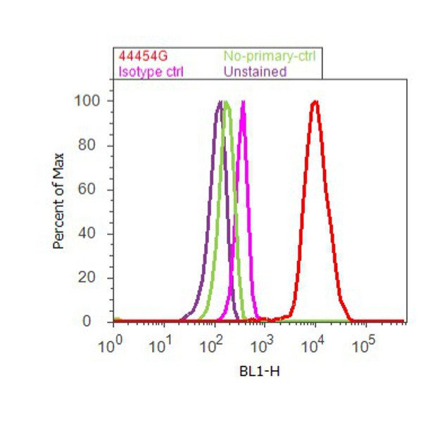
- Flow cytometry analysis of MEK1[pSpS 218/222]/MEK2 [pSpS222/226] was done on A549 cells treated with EGF (200ng/mL, 10 minutes). Cells were fixed with 70% ethanol for 10 minutes, permeabilized with 0.25% Triton™ X-100 for 20 minutes, and blocked with 5% BSA for 30 minutes at room temperature. Cells were labeled withMEK1[pSpS 218/222]/MEK2 [pSpS222/226] Rabbit Polyclonal Antibody (44454G, red histogram) or with rabbit isotype control (pink histogram) at 3-5 ug/million cells in 2.5% BSA. After incubation at room temperature for 2 hours, the cells were labeled with Alexa Fluor® 488 Goat Anti-Rabbit Secondary Antibody (A11008) at a dilution of 1:400 for 30 minutes at room temperature. The representative 10,000 cells were acquired and analyzed for each sample using an Attune® Acoustic Focusing Cytometer. The purple histogram represents unstained control cells and the green histogram represents no-primary-antibody control.