 Explore
 Validate
 Learn
Learn Western blot
Antibody data
- Antibody Data
- Antigen structure
- References [1]
- Comments [0]
- Validations
- Western blot [3]
- Immunocytochemistry [1]
- Immunohistochemistry [1]
- Flow cytometry [1]
Submit
Validation data
Reference
Comment
Report error
- Product number
- 44-458G - Provider product page

- Provider
- Invitrogen Antibodies
- Product name
- Phospho-MEK1 (Thr292) Polyclonal Antibody
- Antibody type
- Polyclonal
- Antigen
- Synthetic peptide
- Reactivity
- Human, Mouse, Rat
- Host
- Rabbit
- Isotype
- IgG
- Vial size
- 100 µL
- Storage
- -20°C
Submitted references The important roles of RET, VEGFR2 and the RAF/MEK/ERK pathway in cancer treatment with sorafenib.
Mao WF, Shao MH, Gao PT, Ma J, Li HJ, Li GL, Han BH, Yuan CG
Acta pharmacologica Sinica 2012 Oct;33(10):1311-8
Acta pharmacologica Sinica 2012 Oct;33(10):1311-8
No comments: Submit comment
Supportive validation
- Submitted by
- Invitrogen Antibodies (provider)
- Main image

- Experimental details
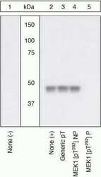
- Peptide Competition Extracts of NIH3T3 cells untreated (lane 1) or treated with 50 ng/mL PDGF for 15 minutes (lanes 2-5) were resolved by SDS-PAGE on a 10% Tris-glycine gel and transferred to PVDF. The membrane was blocked with a 4% BSA-TBST buffer for one hour at room temperature, then incubated with the MEK1 [pT292] antibody (Product # 44-458G) in a 1% BSA-TBST buffer for two hours at room temperature, following prior incubation with: no peptide (1, 2), a generic phosphothreonine-containing peptide (3), the non-phosphopeptide corresponding to the phosphopeptide immunogen (4), or the phosphopeptide immunogen (5). After washing, the membrane was incubated with goat F(ab’)2 anti-rabbit IgG HRP conjugate (Product # ALI4404) and signals were detected using the Pierce SuperSignal™ method. The data show that only the phosphopeptide corresponding to MEK1 [pT292] blocks the antibody signal, demonstrating the specificity of the antibody. The data also show the induction of MEK1 [pT292] phosphorylation by the addition of PDGF to this cell system. In addition, this antibody did not recognize a recombinant MEK1 T292A mutant protein (kindly provided by Dr. Natalie Ahn, University of Colorado), further demonstrating its specificity for MEK1 [pT292] (data not shown).

- Submitted by
- Invitrogen Antibodies (provider)
- Main image
- Experimental details
- Peptide Competition Extracts of NIH3T3 cells untreated (lane 1) or treated with 50 ng/mL PDGF for 15 minutes (lanes 2-5) were resolved by SDS-PAGE on a 10% Tris-glycine gel and transferred to PVDF. The membrane was blocked with a 4% BSA-TBST buffer for one hour at room temperature, then incubated with the MEK1 [pT292] antibody (Product # 44-458G) in a 1% BSA-TBST buffer for two hours at room temperature, following prior incubation with: no peptide (1, 2), a generic phosphothreonine-containing peptide (3), the non-phosphopeptide corresponding to the phosphopeptide immunogen (4), or the phosphopeptide immunogen (5). After washing, the membrane was incubated with goat F(ab’)2 anti-rabbit IgG HRP conjugate (Product # ALI4404) and signals were detected using the Pierce SuperSignal™ method. The data show that only the phosphopeptide corresponding to MEK1 [pT292] blocks the antibody signal, demonstrating the specificity of the antibody. The data also show the induction of MEK1 [pT292] phosphorylation by the addition of PDGF to this cell system. In addition, this antibody did not recognize a recombinant MEK1 T292A mutant protein (kindly provided by Dr. Natalie Ahn, University of Colorado), further demonstrating its specificity for MEK1 [pT292] (data not shown).

- Submitted by
- Invitrogen Antibodies (provider)
- Main image

- Experimental details
- Western blot analysis of MEK1 (pT292) was performed by loading 20 µg of PC-12 (lane1) and NIH\3T3 (lane2) cell lysate using Novex® NuPAGE® 4-12 % Bis-Tris gel (Product # NP0322BOX), XCell SureLock™ Electrophoresis System (Product # EI0002), Novex® Sharp Pre-Stained Protein Standard (LC5800), and iBlot® 2 Dry Blotting System (IB21001). Proteins were transferred to a nitrocellulose membrane and blocked with 5 % skim milk for 1 hour at room temperature. MEK1 (pT292) was detected at ~ 43 kDa using MEK1 (pT292) Rabbit Polyclonal Antibody (Product # 44-244G) at 1:1000 dilution in 5 % skim milk at 4°C overnight on a rocking platform. Goat Anti-Rabbit IgG - HRP Secondary Antibody (G21234) at 1:5000 dilution was used and chemiluminescent detection was performed using Pierce™ ECL Western Blotting Substrate (Product # 32106).

Supportive validation
- Submitted by
- Invitrogen Antibodies (provider)
- Main image

- Experimental details
- Immunofluorescent analysis of Phospho-MEK1 pThr292 Antibody was done on 70% confluent log phase A549 cells. The cells were fixed with 4% paraformaldehyde for 15 minutes, permeabilized with 0.25% Triton™ X-100 for 10 minutes, and blocked with 5% BSA for 1 hour at room temperature. The cells were labeled with Phospho-MEK1 pThr292 Antibody (Product # 44-458G) at 1:250 dilution in 1% BSA and incubated for 3 hours at room temperature and then labeled with Alexa Fluor 488 Goat Anti-Rabbit IgG Secondary Antibody (Product # A-11008) at a dilution of 1:400 for 45 minutes at room temperature (Panel a: green). Nuclei (Panel b: blue) were stained with SlowFade® Gold Antifade Mountant with DAPI (Product # S36938). F-actin (Panel c: red) was stained with Alexa Fluor 594 Phalloidin (Product # A12381). Panel d is a merged image showing nuclear localization. Panel e is a no primary antibody control. The images were captured at 40X magnification.

Supportive validation
- Submitted by
- Invitrogen Antibodies (provider)
- Main image

- Experimental details
- Immunohistochemistry analysis of MEK1 (pT292) showing staining in the cytoplasm of paraffin-embedded human kidney tissue (right) compared to a negative control without primary antibody (left). To expose target proteins, antigen retrieval was performed using 10mM sodium citrate (pH 6.0), microwaved for 8-15 min. Following antigen retrieval, tissues were blocked in 3% H2O2-methanol for 15 min at room temperature, washed with ddH2O and PBS, and then probed with a MEK1 (pT292) polyclonal antibody (Product # 44-458G) diluted in 3% BSA-PBS at a dilution of 1:100 overnight at 4ºC in a humidified chamber. Tissues were washed extensively in PBST and detection was performed using an HRP-conjugated secondary antibody followed by colorimetric detection using a DAB kit. Tissues were counterstained with hematoxylin and dehydrated with ethanol and xylene to prep for mounting.

Supportive validation
- Submitted by
- Invitrogen Antibodies (provider)
- Main image
- Experimental details
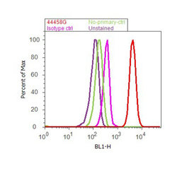
- Flow cytometry analysis of MEK1 [pT292] was done on A549 cells treated with EGF (200ng/mL, 10 minutes). Cells were fixed with 70% ethanol for 10 minutes, permeabilized with 0.25% Triton™ X-100 for 20 minutes, and blocked with 5% BSA for 30 minutes at room temperature. Cells were labeled with MEK1 [pT292] Rabbit Polyclonal Antibody (44458G, red histogram) or with rabbit isotype control (pink histogram) at 3-5 ug/million cells in 2.5% BSA. After incubation at room temperature for 2 hours, the cells were labeled with Alexa Fluor® 488 Goat Anti-Rabbit Secondary Antibody (A11008) at a dilution of 1:400 for 30 minutes at room temperature. The representative 10,000 cells were acquired and analyzed for each sample using an Attune® Acoustic Focusing Cytometer. The purple histogram represents unstained control cells and the green histogram represents no-primary-antibody control.
