Explore
 Validate
 Learn
Learn Western blot
 Immunocytochemistry
Antibody data
- Antibody Data
- Antigen structure
- References [3]
- Comments [0]
- Validations
- Immunocytochemistry [3]
- Immunohistochemistry [2]
- Flow cytometry [1]
- Other assay [2]
Submit
Validation data
Reference
Comment
Report error
- Product number
- 44-460G - Provider product page

- Provider
- Invitrogen Antibodies
- Product name
- Phospho-MEK1 (Ser298) Polyclonal Antibody
- Antibody type
- Polyclonal
- Antigen
- Synthetic peptide
- Reactivity
- Human, Mouse, Rat
- Host
- Rabbit
- Isotype
- IgG
- Vial size
- 100 μL
- Storage
- -20°C
Submitted references Group I Paks are essential for epithelial- mesenchymal transition in an Apc-driven model of colorectal cancer.
Group I Paks as therapeutic targets in NF2-deficient meningioma.
The important roles of RET, VEGFR2 and the RAF/MEK/ERK pathway in cancer treatment with sorafenib.
Chow HY, Dong B, Valencia CA, Zeng CT, Koch JN, Prudnikova TY, Chernoff J
Nature communications 2018 Aug 27;9(1):3473
Nature communications 2018 Aug 27;9(1):3473
Group I Paks as therapeutic targets in NF2-deficient meningioma.
Chow HY, Dong B, Duron SG, Campbell DA, Ong CC, Hoeflich KP, Chang LS, Welling DB, Yang ZJ, Chernoff J
Oncotarget 2015 Feb 10;6(4):1981-94
Oncotarget 2015 Feb 10;6(4):1981-94
The important roles of RET, VEGFR2 and the RAF/MEK/ERK pathway in cancer treatment with sorafenib.
Mao WF, Shao MH, Gao PT, Ma J, Li HJ, Li GL, Han BH, Yuan CG
Acta pharmacologica Sinica 2012 Oct;33(10):1311-8
Acta pharmacologica Sinica 2012 Oct;33(10):1311-8
No comments: Submit comment
Supportive validation
- Submitted by
- Invitrogen Antibodies (provider)
- Main image
- Experimental details
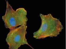
- MEK1 (pS298) phosphospecific antibody. A549 cells plated on fibronectin, serum starved overnight then stimulated with serum for 1 hour. Green = MEK1 (pS298), red = actin, blue = Hoechst 33342.

- Submitted by
- Invitrogen Antibodies (provider)
- Main image

- Experimental details
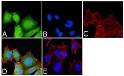
- Immunofluorescent analysis of Phospho-MEK1 pSer298 Antibody was done on 70% confluent log phase A549 cells. The cells were fixed with 4% paraformaldehyde for 15 minutes, permeabilized with 0.25% Triton™ X-100 for 10 minutes, and blocked with 5% BSA for 1 hour at room temperature. The cells were labeled with Phospho-MEK1 pSer298Antibody (Product # 44-460G) at 1:250 dilution in 1% BSA and incubated for 3 hours at room temperature and then labeled with Alexa Fluor 488 Goat Anti-Rabbit IgG Secondary Antibody (Product # A-11008) at a dilution of 1:400 for 45 minutes at room temperature (Panel a: green). Nuclei (Panel b: blue) were stained with SlowFade® Gold Antifade Mountant with DAPI (Product # S36938). F-actin (Panel c: red) was stained with Alexa Fluor 594 Phalloidin (Product # A12381). Panel d is a merged image showing cytoplasmic and nuclear localization. Panel e is a no primary antibody control. The images were captured at 40X magnification.

- Submitted by
- Invitrogen Antibodies (provider)
- Main image
- Experimental details
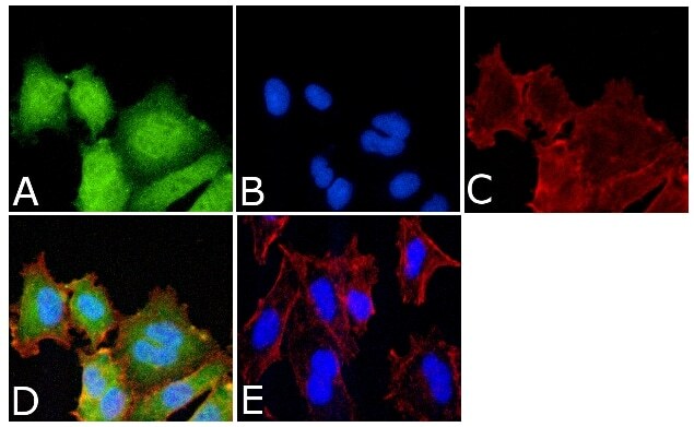
- Immunofluorescent analysis of Phospho-MEK1 pSer298 Antibody was done on 70% confluent log phase A549 cells. The cells were fixed with 4% paraformaldehyde for 15 minutes, permeabilized with 0.25% Triton™ X-100 for 10 minutes, and blocked with 5% BSA for 1 hour at room temperature. The cells were labeled with Phospho-MEK1 pSer298Antibody (Product # 44-460G) at 1:250 dilution in 1% BSA and incubated for 3 hours at room temperature and then labeled with Alexa Fluor 488 Goat Anti-Rabbit IgG Secondary Antibody (Product # A-11008) at a dilution of 1:400 for 45 minutes at room temperature (Panel a: green). Nuclei (Panel b: blue) were stained with SlowFade® Gold Antifade Mountant with DAPI (Product # S36938). F-actin (Panel c: red) was stained with Alexa Fluor 594 Phalloidin (Product # A12381). Panel d is a merged image showing cytoplasmic and nuclear localization. Panel e is a no primary antibody control. The images were captured at 40X magnification.
Supportive validation
- Submitted by
- Invitrogen Antibodies (provider)
- Main image
- Experimental details
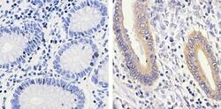
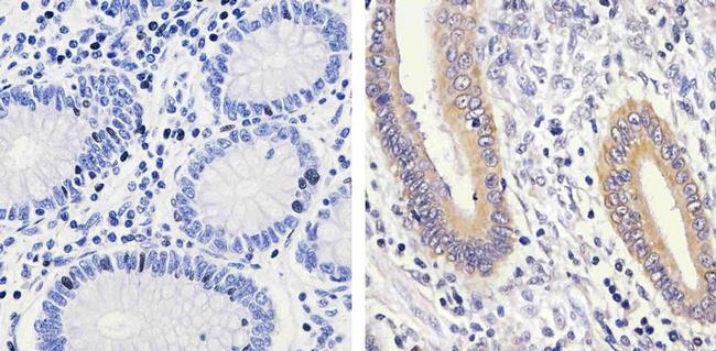
- Immunohistochemistry analysis of MEK1 (pS298) showing staining in the cytoplasm of paraffin-embedded human colon carcinoma tissue (right) compared to a negative control without primary antibody (left). To expose target proteins, antigen retrieval was performed using 10mM sodium citrate (pH 6.0), microwaved for 8-15 min. Following antigen retrieval, tissues were blocked in 3% H2O2-methanol for 15 min at room temperature, washed with ddH2O and PBS, and then probed with a MEK1 (pS298) polyclonal antibody (Product # 44-460G) diluted in 3% BSA-PBS at a dilution of 1:100 overnight at 4ºC in a humidified chamber. Tissues were washed extensively in PBST and detection was performed using an HRP-conjugated secondary antibody followed by colorimetric detection using a DAB kit. Tissues were counterstained with hematoxylin and dehydrated with ethanol and xylene to prep for mounting.
- Submitted by
- Invitrogen Antibodies (provider)
- Main image
- Experimental details
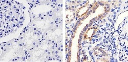
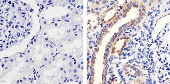
- Immunohistochemistry analysis of MEK1 (pS298) showing staining in the cytoplasm of paraffin-embedded human kidney tissue (right) compared to a negative control without primary antibody (left). To expose target proteins, antigen retrieval was performed using 10mM sodium citrate (pH 6.0), microwaved for 8-15 min. Following antigen retrieval, tissues were blocked in 3% H2O2-methanol for 15 min at room temperature, washed with ddH2O and PBS, and then probed with a MEK1 (pS298) polyclonal antibody (Product # 44-460G) diluted in 3% BSA-PBS at a dilution of 1:100 overnight at 4ºC in a humidified chamber. Tissues were washed extensively in PBST and detection was performed using an HRP-conjugated secondary antibody followed by colorimetric detection using a DAB kit. Tissues were counterstained with hematoxylin and dehydrated with ethanol and xylene to prep for mounting.
Supportive validation
- Submitted by
- Invitrogen Antibodies (provider)
- Main image
- Experimental details
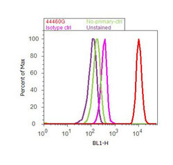
- Flow cytometry analysis of MEK1 [pS298] was done on A549 cells treated with EGF (200ng/mL, 10 minutes). Cells were fixed with 70% ethanol for 10 minutes, permeabilized with 0.25% Triton™ X-100 for 20 minutes, and blocked with 5% BSA for 30 minutes at room temperature. Cells were labeled with MEK1 [pS298] Rabbit Polyclonal Antibody (44460G, red histogram) or with rabbit isotype control (pink histogram) at 3-5 ug/million cells in 2.5% BSA. After incubation at room temperature for 2 hours, the cells were labeled with Alexa Fluor® 488 Goat Anti-Rabbit Secondary Antibody (A11008) at a dilution of 1:400 for 30 minutes at room temperature. The representative 10,000 cells were acquired and analyzed for each sample using an Attune® Acoustic Focusing Cytometer. The purple histogram represents unstained control cells and the green histogram represents no-primary-antibody control.
Supportive validation
- Submitted by
- Invitrogen Antibodies (provider)
- Main image
- Experimental details
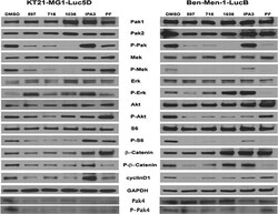
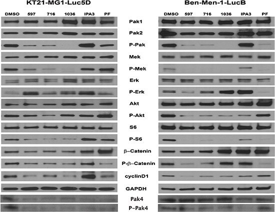
- Figure 4 The effect of Pak inhibitors on Erk and Akt-S6 signaling pathways KT21 or Ben-Men cells were treated with inhibitors for 72 hours as described in Figure 2 . Following SDS/PAGE and transfer to PVDF membranes, expression levels of Pak, Mek, Erk, Akt, S6, and beta-Catenin were assessed by immunoblot using total and phospho-specific antibodies. GADPH was used as loading control.
- Submitted by
- Invitrogen Antibodies (provider)
- Main image
- Experimental details
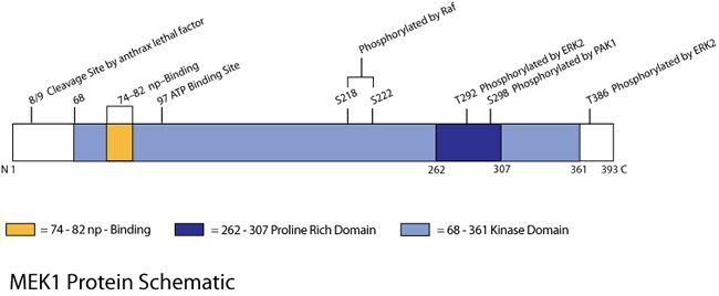
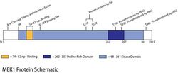
- MEK Protein Schematic-Originally cloned from mouse cDNA, MEK1 (MAP kinase kinase-1) is a 43.5 kDa protein which phosphorylates ERK1&2 in the MAP kinase signaling pathway. Soon after the discovery of MEK1, MKK1 (the human homologue) was cloned. Like ERK1&2, MEK1 is primarily involved in cell growth and differentiation through its regulation of the cell cycle. G-proteins such as MEKK (MEK kinase) or Raf phosphorylate MEK1/2 at serine residues in order to activate the enzymatic activity of the protein. In 1999, Sebolt-Leopold, found that MEK1/2 overexpression was linked to cellular transformation, which sparked investigation of possible therapeutic potentials for MEK1/2 inhibitors. Several inhibitors including U-0126 and PD98059 were developed and have shown promising results as therapies for cancer and other diseases.