Explore
 Validate
 Learn
Learn Western blot
 Immunocytochemistry
Antibody data
- Antibody Data
- Antigen structure
- References [1]
- Comments [0]
- Validations
- Western blot [2]
- Immunohistochemistry [2]
Submit
Validation data
Reference
Comment
Report error
- Product number
- NBP1-87790 - Provider product page

- Provider
- Novus Biologicals
- Proper citation
- Novus Cat#NBP1-87790, RRID:AB_11023812
- Product name
- Rabbit Polyclonal MEK1 Antibody
- Antibody type
- Polyclonal
- Description
- Immunogen affinity purified. Specificity of human, rat MEK1 antibody verified on a Protein Array containing target protein plus 383 other non-specific proteins.
- Reactivity
- Human, Rat
- Host
- Rabbit
- Isotype
- IgG
- Vial size
- 0.1 ml
- Storage
- Store at 4C short term. Aliquot and store at -20C long term. Avoid freeze-thaw cycles.
Submitted references MEK1 is associated with carboplatin resistance and is a prognostic biomarker in epithelial ovarian cancer.
Pénzváltó Z, Lánczky A, Lénárt J, Meggyesházi N, Krenács T, Szoboszlai N, Denkert C, Pete I, Győrffy B
BMC cancer 2014 Nov 18;14:837
BMC cancer 2014 Nov 18;14:837
No comments: Submit comment
Supportive validation
- Submitted by
- Novus Biologicals (provider)
- Main image
- Experimental details
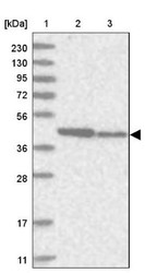
- Western Blot: MEK1 Antibody [NBP1-87790] - Lane 1: Marker [kDa] 230, 130, 95, 72, 56, 36, 28, 17, 11. Lane 2: Human cell line RT-4. Lane 3: Human cell line U-251MG sp
- Submitted by
- Novus Biologicals (provider)
- Main image
- Experimental details
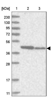
- Western Blot: MEK1 Antibody [NBP1-87790] - Lane 1: NIH-3T3 cell lysate (Mouse embryonic fibroblast cells). Lane 2: NBT-II cell lysate (Rat Wistar bladder tumor cells).

Supportive validation
- Submitted by
- Novus Biologicals (provider)
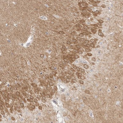
- Main image
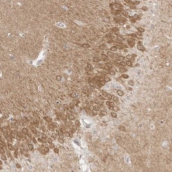
- Experimental details
- Immunohistochemistry-Paraffin: MEK1 Antibody [NBP1-87790] - Staining of human hippocampus shows moderate cytoplasmic positivity in neuronal cells.
- Submitted by
- Novus Biologicals (provider)
- Main image

- Experimental details
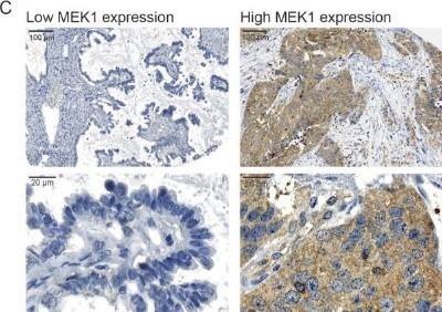
- Immunohistochemistry: MEK1 Antibody [NBP1-87790] - Correlation between MEK1 expression and survival after platinum treatment in EOC patients. Representative images of immunohistochemistry, low and high expression of MEK1 at low and high magnifications. Image collected and cropped by CiteAb from the following publication (http://bmccancer.biomedcentral.com/articles/10.1186/1471-2407-14-837), licensed under a CC-BY licence.