Explore
 Validate
 Learn
Learn Western blot
Antibody data
- Antibody Data
- Antigen structure
- References [2]
- Comments [0]
- Validations
- Western blot [3]
- Immunocytochemistry [1]
- Immunohistochemistry [1]
Submit
Validation data
Reference
Comment
Report error
- Product number
- PA5-31917 - Provider product page

- Provider
- Invitrogen Antibodies
- Product name
- MEK1/MEK2 Polyclonal Antibody
- Antibody type
- Polyclonal
- Antigen
- Recombinant protein fragment
- Description
- Recommended positive controls: 293T, NIH-3T3, JC.
- Concentration
- 1 mg/mL
Submitted references Oncogenic RAS drives the CRAF-dependent extracellular vesicle uptake mechanism coupled with metastasis.
Fat1 deletion promotes hybrid EMT state, tumour stemness and metastasis.
Choi D, Montermini L, Meehan B, Lazaris A, Metrakos P, Rak J
Journal of extracellular vesicles 2021 Jun;10(8):e12091
Journal of extracellular vesicles 2021 Jun;10(8):e12091
Fat1 deletion promotes hybrid EMT state, tumour stemness and metastasis.
Pastushenko I, Mauri F, Song Y, de Cock F, Meeusen B, Swedlund B, Impens F, Van Haver D, Opitz M, Thery M, Bareche Y, Lapouge G, Vermeersch M, Van Eycke YR, Balsat C, Decaestecker C, Sokolow Y, Hassid S, Perez-Bustillo A, Agreda-Moreno B, Rios-Buceta L, Jaen P, Redondo P, Sieira-Gil R, Millan-Cayetano JF, Sanmatrtin O, D'Haene N, Moers V, Rozzi M, Blondeau J, Lemaire S, Scozzaro S, Janssens V, De Troya M, Dubois C, Pérez-Morga D, Salmon I, Sotiriou C, Helmbacher F, Blanpain C
Nature 2021 Jan;589(7842):448-455
Nature 2021 Jan;589(7842):448-455
No comments: Submit comment
Supportive validation
- Submitted by
- Invitrogen Antibodies (provider)
- Main image
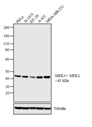
- Experimental details
- Western blot analysis was performed on whole cell extracts (30 µg lysate) of HeLa (Lane 1), H-1975 (Lane 2), HT-29 (Lane 3), A-431 (Lane 4) and MDA-MB-231 (Lane 5). The blot was probed with Anti-MEK1/MEK2 Rabbit Polyclonal Antibody (Product # PA5-31917, 1 µg/mL) and detected by chemiluminescence using Goat anti-Rabbit IgG (H+L) Superclonal™ Secondary Antibody, HRP conjugate (Product # A27036, 0.25 µg/mL, 1:4000 dilution). A 45 kDa band corresponding to MEK1/MEK2 was detected across all cell lines tested. Known quantity of protein samples were electrophoresed using Novex® NuPAGE® 4-12 % Bis-Tris gel (Product # NP0321BOX), XCell SureLock™ Electrophoresis System (Product # EI0002) and Novex® Sharp Pre-Stained Protein Standard (Product # LC5800). Resolved proteins were then transferred onto a nitrocellulose membrane with iBlot® 2 Dry Blotting System (Product # IB21001). The membrane was probed with the relevant primary and secondary Antibody following blocking with 5 % skimmed milk. Chemiluminescent detection was performed using Pierce™ ECL Western Blotting Substrate (Product # 32106).

- Submitted by
- Invitrogen Antibodies (provider)
- Main image
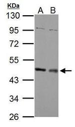
- Experimental details
- Western Blot using MEK1/MEK2 Polyclonal Antibody (Product # PA5-31917). Sample (30 µg of whole cell lysate). Lane A: NIH-3T3. Lane B: JC. 10% SDS PAGE. MEK1/MEK2 Polyclonal Antibody (Product # PA5-31917) diluted at 1:2,000.
- Submitted by
- Invitrogen Antibodies (provider)
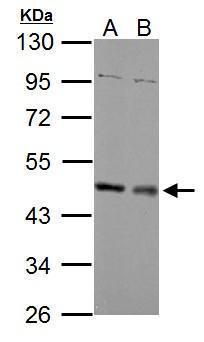
- Main image

- Experimental details
- Western Blot using MEK1/MEK2 Polyclonal Antibody (Product # PA5-31917). Sample (30 µg of whole cell lysate). Lane A: 293T. 10% SDS PAGE. MEK1/MEK2 Polyclonal Antibody (Product # PA5-31917) diluted at 1:3,000.

Supportive validation
- Submitted by
- Invitrogen Antibodies (provider)
- Main image
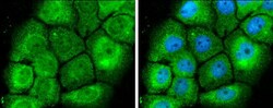
- Experimental details
- Immunocytochemistry-Immunofluorescence analysis of MEK1/MEK2 was performed in A431 cells fixed in 4% paraformaldehyde at RT for 15 min. Green: MEK1/MEK2 Polyclonal Antibody (Product # PA5-31917) diluted at 1:500. Blue: Hoechst 33342 staining.

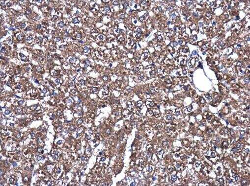
Supportive validation
- Submitted by
- Invitrogen Antibodies (provider)
- Main image
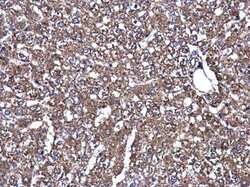
- Experimental details
- Immunohistochemistry (Paraffin) analysis of MEK1/MEK2 was performed in paraffin-embedded rat liver tissue using MEK1/MEK2 Polyclonal Antibody (Product # PA5-31917) at a dilution of 1:500.