Explore
 Validate
 Learn
Learn Western blot
Antibody data
- Antibody Data
- Antigen structure
- References [3]
- Comments [0]
- Validations
- Western blot [4]
- Immunocytochemistry [1]
- Immunohistochemistry [1]
Submit
Validation data
Reference
Comment
Report error
- Product number
- GTX103174 - Provider product page

- Provider
- GeneTex
- Proper citation
- GeneTex Cat#GTX103174, RRID:AB_1951777
- Product name
- p70 S6K antibody
- Antibody type
- Polyclonal
- Reactivity
- Human, Mouse, Rat
- Host
- Rabbit
- Storage
- Keep as concentrated solution. Aliquot and store at -20°C or below. Avoid multiple freeze-thaw cycles.
Submitted references RagA, an mTORC1 activator, interacts with a hedgehog signaling protein, WDR35/IFT121.
Rhapontigenin inhibits TGF-β-mediated epithelial‑mesenchymal transition via the PI3K/AKT/mTOR pathway and is not associated with HIF-1α degradation.
A gene expression signature-based approach reveals the mechanisms of action of the Chinese herbal medicine berberine.
Sekiguchi T, Furuno N, Ishii T, Hirose E, Sekiguchi F, Wang Y, Kobayashi H
Genes to cells : devoted to molecular & cellular mechanisms 2019 Feb;24(2):151-161
Genes to cells : devoted to molecular & cellular mechanisms 2019 Feb;24(2):151-161
Rhapontigenin inhibits TGF-β-mediated epithelial‑mesenchymal transition via the PI3K/AKT/mTOR pathway and is not associated with HIF-1α degradation.
Yeh YH, Wang SW, Yeh YC, Hsiao HF, Li TK
Oncology reports 2016 May;35(5):2887-95
Oncology reports 2016 May;35(5):2887-95
A gene expression signature-based approach reveals the mechanisms of action of the Chinese herbal medicine berberine.
Lee KH, Lo HL, Tang WC, Hsiao HH, Yang PM
Scientific reports 2014 Sep 17;4:6394
Scientific reports 2014 Sep 17;4:6394
No comments: Submit comment
Supportive validation
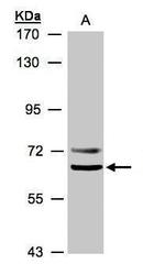
- Submitted by
- GeneTex (provider)
- Main image
- Experimental details
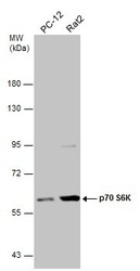
- Sample (30 μg of whole cell lysate) A: NIH-3T3 7.5% SDS PAGE GTX103174 diluted at 1:10000

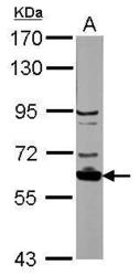
- Submitted by
- GeneTex (provider)
- Main image
- Experimental details
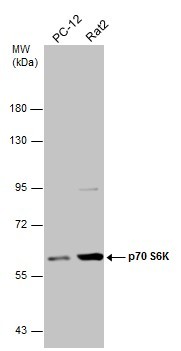
- Sample(30 ug whole cell lysate)A:293T7.5% SDS PAGEGTX103174 diluted at 1:500
- Validation comment
- WB
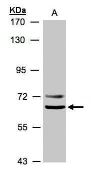
- Submitted by
- GeneTex (provider)
- Main image
- Experimental details
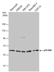
- Various whole cell extracts (30 μg) were separated by 7.5% SDS-PAGE, and the membrane was blotted with p70 S6K antibody (GTX103174) diluted at 1:50000.
- Submitted by
- GeneTex (provider)
- Main image
- Experimental details
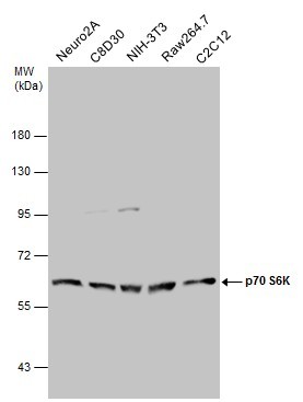
- Various whole cell extracts (30 μg) were separated by 7.5% SDS-PAGE, and the membrane was blotted with p70 S6K antibody (GTX103174) diluted at 1:50000.
Supportive validation
- Submitted by
- GeneTex (provider)
- Main image
- Experimental details
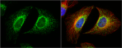
- p70 S6K antibody detects p70 S6K protein at mitochondria by immunofluorescent analysis.Sample: HeLa cells were fixed in 4% paraformaldehyde at RT for 15 min.Green: p70 S6K protein stained by p70 S6K antibody (GTX103174) diluted at 1:1000.Red: alpha Tubulin, a cytoskeleton marker, stained by alpha Tubulin antibody [B-5-1-2] (GTX11304) diluted at 1:10000.Blue: Hoechst 33342 staining.

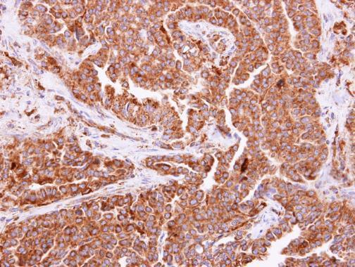
Supportive validation
- Submitted by
- GeneTex (provider)
- Main image
- Experimental details
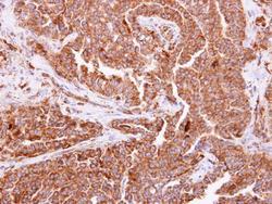
- p70 S6K antibody detects p70 S6K protein at cytoplasm on human lung carcinoma by immunohistochemical analysis. Sample: Paraffin-embedded lung carcinoma. p70 S6K antibody (GTX103174) dilution: 1:500.