Explore
 Validate
 Learn
Learn Western blot
Antibody data
- Antibody Data
- Antigen structure
- References [2]
- Comments [0]
- Validations
- Western blot [2]
- Immunohistochemistry [2]
- Flow cytometry [1]
Submit
Validation data
Reference
Comment
Report error
- Product number
- AF8962 - Provider product page

- Provider
- R&D Systems
- Product name
- Human/Mouse/Rat p70 S6 Kinase Antibody
- Antibody type
- Polyclonal
- Description
- Immunogen affinity purified. Detects human, mouse, and rat p70 S6K and p85 S6K (also known as p70 S6K alpha I), an isoform with 23 extra residues at the N-terminus. Reactivity with beta isoforms of p70 S6K is unknown.
- Reactivity
- Human, Mouse, Rat
- Host
- Rabbit
- Conjugate
- Unconjugated
- Antigen sequence
M60725- Isotype
- IgG
- Vial size
- 100 ug
- Concentration
- LYOPH
- Storage
- Use a manual defrost freezer and avoid repeated freeze-thaw cycles. 12 months from date of receipt, -20 to -70 °C as supplied. 1 month, 2 to 8 °C under sterile conditions after reconstitution. 6 months, -20 to -70 °C under sterile conditions after reconstitution.
Submitted references Mild MPP(+) exposure-induced glucose starvation enhances autophagosome synthesis and impairs its degradation.
The role of autophagy in cardiomyocytes in the basal state and in response to hemodynamic stress.
Sakamoto S, Miyara M, Sanoh S, Ohta S, Kotake Y
Scientific reports 2017 Apr 26;7:46668
Scientific reports 2017 Apr 26;7:46668
The role of autophagy in cardiomyocytes in the basal state and in response to hemodynamic stress.
Nakai A, Yamaguchi O, Takeda T, Higuchi Y, Hikoso S, Taniike M, Omiya S, Mizote I, Matsumura Y, Asahi M, Nishida K, Hori M, Mizushima N, Otsu K
Nature medicine 2007 May;13(5):619-24
Nature medicine 2007 May;13(5):619-24
No comments: Submit comment
Supportive validation
- Submitted by
- R&D Systems (provider)
- Main image
- Experimental details
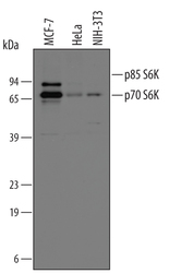
- Detection of Human and Mouse p70 S6 Kinase by Western Blot. Western blot shows lysates of MCF-7 human breast cancer cell line, HeLa human cervical epithelial carcinoma cell line, and NIH-3T3 mouse embryonic fibroblast cell line. PVDF membrane was probed with 0.2 µg/mL of Rabbit Anti-Human/Mouse/Rat p70 S6 Kinase Antigen Affinity-purified Polyclonal Antibody (Catalog # AF8962) followed by HRP-conjugated Anti-Rabbit IgG Secondary Antibody (Catalog # HAF008). A specific band was detected for p70 S6 Kinase at approximately 70 and 90 kDa (as indicated). This experiment was conducted under reducing conditions and using Immunoblot Buffer Group 1.
- Submitted by
- R&D Systems (provider)
- Main image
- Experimental details
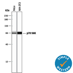
- Detection of p70 S6 Kinase by Simple WesternTM. Simple Western lane view shows lysates of HeLa human cervical epithelial carcinoma cell line and NIH-3T3 mouse embryonic fibroblast cell line, loaded at 0.2 mg/mL. A specific band was detected for p70 S6 Kinase at approximately 66 kDa (as indicated) using 2 µg/mL of Rabbit Anti-Human/Mouse/Rat p70 S6 Kinase Antigen Affinity-purified Polyclonal Antibody (Catalog # AF8962). This experiment was conducted under reducing conditions and using the 12-230 kDa separation system.

Supportive validation
- Submitted by
- R&D Systems (provider)
- Main image
- Experimental details
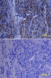
- p70 S6 Kinase in Human Breast Cancer Tissue. p70 S6 Kinase was detected in immersion fixed paraffin-embedded sections of human breast cancer tissue using Rabbit Anti-Human/Mouse/Rat p70 S6 Kinase Antigen Affinity-purified Polyclonal Antibody (Catalog # AF8962) at 15 µg/mL overnight at 4 °C. Tissue was stained using the Anti-Rabbit HRP-DAB Cell & Tissue Staining Kit (brown; Catalog # CTS005) and counterstained with hematoxylin (blue). Lower panel shows a lack of labeling if primary antibodies are omitted and tissue is stained only with secondary antibody followed by incubation with detection reagents. View our protocol for Chromogenic IHC Staining of Paraffin-embedded Tissue Sections.

- Submitted by
- R&D Systems (provider)
- Main image

- Experimental details
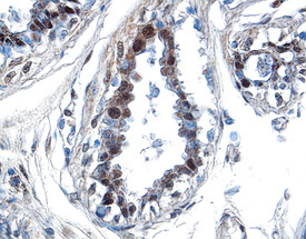
- p70 S6 Kinase in Human Breast Cancer Tissue. p70 S6 Kinase was detected in immersion fixed paraffin-embedded sections of human breast cancer tissue using Rabbit Anti-Human/Mouse/Rat p70 S6 Kinase Antigen Affinity-purified Polyclonal Antibody (Catalog # AF8962) at 1.7 µg/mL overnight at 4 °C. Tissue was stained using the Anti-Rabbit HRP-DAB Cell & Tissue Staining Kit (brown; Catalog # CTS005) and counterstained with hematoxylin (blue). Specific labeling was localized to the nuclei of epithelial cells. View our protocol for Chromogenic IHC Staining of Paraffin-embedded Tissue Sections.
Supportive validation
- Submitted by
- R&D Systems (provider)
- Main image

- Experimental details
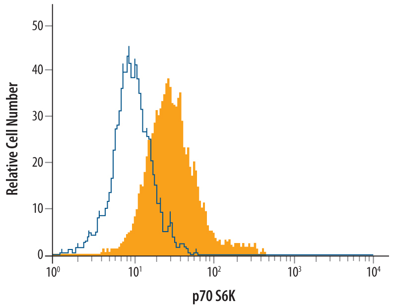
- Detection of p70 S6 Kinase in HeLa Human Cell Line by Flow Cytometry. HeLa human cervical epithelial carcinoma cell line was stained with Rabbit Anti-Human/Mouse/Rat p70 S6 Kinase Antigen Affinity-purified Polyclonal Antibody (Catalog # AF8962, filled histogram) or control antibody (Catalog # AB-105-C, open histogram), followed by Phycoerythrin-conjugated Anti-Rabbit IgG Secondary Antibody (Catalog # F0110). To facilitate intracellular staining, cells were fixed with paraformaldehyde and permeabilized with methanol.