 Explore
 Validate
 Learn
Learn Western blot
 ELISA
ELISAAntibody data
- Antibody Data
- Antigen structure
- References [7]
- Comments [0]
- Validations
- Western blot [2]
Submit
Validation data
Reference
Comment
Report error
- Product number
- ABIN1741708 - Provider product page

- Provider
- antibodies-online
- Product name
- anti-Poly (ADP-Ribose) Polymerase 1 (PARP1) antibody
- Antibody type
- Polyclonal
- Antigen
- PARP1, human
- Description
- Immunofractionation
- Reactivity
- Human, Mouse, Rat
- Host
- Chicken/Avian
- Isotype
- IgY
- Vial size
- 1 mg
- Concentration
- 1 mg/mL
- Storage
- Stable for 1 year from date of shipment when stored at -20°C or -70°C. Stable for at least 1 month at 4°C.
- Handling
- Avoid freeze/thaw cycles.
Submitted references Transcriptional control by PARP-1: chromatin modulation, enhancer-binding, coregulation, and insulation.
Poly ADP-ribose polymerase-1: an international molecule of mystery.
The diverse biological roles of mammalian PARPS, a small but powerful family of poly-ADP-ribose polymerases.
Characterization of the necrotic cleavage of poly(ADP-ribose) polymerase (PARP-1): implication of lysosomal proteases.
Characterization of antibodies specific for the caspase cleavage site on poly(ADP-ribose) polymerase: specific detection of apoptotic fragments and mapping of the necrotic fragments of poly(ADP-ribose) polymerase.
Characterization of anti-peptide antibodies directed towards the automodification domain and apoptotic fragment of poly (ADP-ribose) polymerase.
Cleavage of poly(ADP-ribose) polymerase by a proteinase with properties like ICE.
Kraus WL
Current opinion in cell biology 2008 Jun;20(3):294-302
Current opinion in cell biology 2008 Jun;20(3):294-302
Poly ADP-ribose polymerase-1: an international molecule of mystery.
Woodhouse BC, Dianov GL
DNA repair 2008 Jul 1;7(7):1077-86
DNA repair 2008 Jul 1;7(7):1077-86
The diverse biological roles of mammalian PARPS, a small but powerful family of poly-ADP-ribose polymerases.
Hassa PO, Hottiger MO
Frontiers in bioscience : a journal and virtual library 2008 Jan 1;13:3046-82
Frontiers in bioscience : a journal and virtual library 2008 Jan 1;13:3046-82
Characterization of the necrotic cleavage of poly(ADP-ribose) polymerase (PARP-1): implication of lysosomal proteases.
Gobeil S, Boucher CC, Nadeau D, Poirier GG
Cell death and differentiation 2001 Jun;8(6):588-94
Cell death and differentiation 2001 Jun;8(6):588-94
Characterization of antibodies specific for the caspase cleavage site on poly(ADP-ribose) polymerase: specific detection of apoptotic fragments and mapping of the necrotic fragments of poly(ADP-ribose) polymerase.
Sallmann FR, Bourassa S, Saint-Cyr J, Poirier GG
Biochemistry and cell biology = Biochimie et biologie cellulaire 1997;75(4):451-6
Biochemistry and cell biology = Biochimie et biologie cellulaire 1997;75(4):451-6
Characterization of anti-peptide antibodies directed towards the automodification domain and apoptotic fragment of poly (ADP-ribose) polymerase.
Duriez PJ, Desnoyers S, Hoflack JC, Shah GM, Morelle B, Bourassa S, Poirier GG, Talbot B
Biochimica et biophysica acta 1997 Feb 11;1334(1):65-72
Biochimica et biophysica acta 1997 Feb 11;1334(1):65-72
Cleavage of poly(ADP-ribose) polymerase by a proteinase with properties like ICE.
Lazebnik YA, Kaufmann SH, Desnoyers S, Poirier GG, Earnshaw WC
Nature 1994 Sep 22;371(6495):346-7
Nature 1994 Sep 22;371(6495):346-7
No comments: Submit comment
Supportive validation
- Submitted by
- antibodies-online (provider)
- Main image

- Experimental details
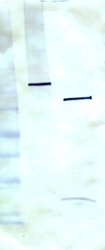
- Western blot using anti-PARP1. Lane 1, control Jurkat cell lysate (5 ?g); Lane 2, apoptotic Jurkat cell lysate, induced with camptothecin for 4 hr. Arrows point to intact 113kDa PARP and apoptosis-induced 89kDa cleavage fragment. Note other smaller apoptosisinduced PARP fragments. Primary antibody concentration was 1 ?g/mL, secondary GAC-HRP was 1/2000, followed by color development with TMB ~5 min.

- Submitted by
- antibodies-online (provider)
- Main image
- Experimental details
- Western blot using anti-PARP1, clone C-2-10. Lane 1, control Jurkat cell lysate; Lane 2, apoptotic Jurkat cell lysate. This monoclonal antibody recognizes only the intact 113kDa and apoptotic 89kDa fragment. This blot is shown for comparison purposes.
